Вы абсолютно правы! Это классическая гемангиома печени. Остальные фазы не были опубликованы намеренно. По последним данным; для точной диагностики гемангиом печени, вполне достаточно исследования в артериальную фазу; Вы спросите почему? На эту тему я как раз и предлагаю поговорить.
Марио я с Вами соглашусь, даже и своего скромного опыта замечал, что заряжая многофазовое исследование гемангиому узнаешь сразу на артериальной, а все отсальные фазы идут без диагностической ценности.
КТ/МРТ исследования печени требуют чётких и обоснованных протоколов, невозможно все пациентам выполнять ,нативные, ранние/поздние артериальные, портальные и отсроченные фазы. Это трудоёмко, весьма затратно о несёт серьёзную лучевую нагрузку для пациента. Было отмечено, что образования печен с периферическим усилением в артериальную фазу, имеют два типа контрастирования (lesions with peripheral contrast attenuations at arterial phase):
1. contineous peripheral ring-like enhancement: по типу замкнутого кольца: метастазы метастазы и абсцессы (также грибковые поражения, например кандидоз). 2. non-contineous peripheral puddle-like enhancement: по типу не-замкнутого периферического усиления с отдельными "лакунами", иногда с центрипетальными "языками" (centripetal contarst extensions); характерных исключительно для гемангиом.
Вывод: если идёт КТ исследование брюшной полости, и вся желаемая диагностическая информация была получена на поздней артериальной фазе, при обнаружении характерной картины гемангиомы печени, можно остановить исследование и не выполнять отсроченные венозные фазы, экономя деньги, амортизацию аппарата и конечно заботясь о лучевой безопасности пациента.
Марио. Вопрос! как быть с мизерными гемангиомами, они в артериальную фазу "вспыхивают" одномоментно, как же тут применить тип контрастирования?. У меня реально существует проблема в диф.диагностики малых образований, толи это гемангиомы, толи гиперваскулярные метастазы (были случаи), причем я ориентировался именно на отсроченные фазы, известно, что гемангиомы выравниваются с плотностью печени, а метастазы нет+ быстро "вымывают" контраст.
Интересный вопрос, в таких случаях (множественные гиперваскулярные образования), отсроченную фазу делать необходимо, исключить метастазы является первейшей задачей. Таk что всё Вы делаете правильно. И заметьте, при мелких множественных гемангиомах, они могут контрастироваться (как Вы правильно заметили "вспыхивать"), полностью, так что говорить о "периферическом типе усиления с лакунами контраста" в таких случаях нельзя, следовательно и правило на сработает.
Еще один вопрос. Марио у Вас были ситуации например: на КТ данных за гемангиому нет, а при верификации она есть!?. К тому что все ли гемангиомы, именно ВСЕ и ВСЕГДА имеют патогномоничную картину контрастирования. Бывало что я встречался с такими заключениями как атипичные, гиповаскулярные гемангиомы. Но классификация не подразумевает таких видов гемангиом, и совершенно не понятно на каких признаках выносятся такие заключения.
Вы задали очень сложный вопрос. Мы все прекрасно знаем; что медицина это не математика, всегда будет 5-10% патологии, которая не подходить под классическое описание. Термин атипическая гемангиома печени; используют в УЗИ диагностике, имеется ввиду такие гемангиомы, которые имеет не-классическую (гиперэхогенную),атипическую презентацию. Как правило они в разной степени гипоэхогенны, особенно в центральной части (могут быть гетерогенными) с гипрэхогенным ободком. Такая презентация объясняется последствиями центрального геморрагического некрозом, формированием рубцов или миксоматозными изменениями. Некоторые авторы заявляют, что процент таких атипичных гемангиом достигает 40% (!) (Moody and Wilson 1993).
В отношении КТ и МРТ; более распространён термин: гемангиомы с атипическим контрастированием; как правило такая атипия объясняется изменениями в окружающей печёночной ткани, например наличие фиброзных изменений за счёт активного вирусного гепатита С: посмотрите эту статью (можно скачать .PDF):
http://www.springerlink.com/content/n11n762475112635/ Masakatsu Tsurusaki1, 3 Contact Information, Ryota Kawasaki1, Masato Yamaguchi1, Koji Sugimoto1, Takumi Fukumoto2, Yonson Ku2 and Kazuro Sugimura1 (1) Department of Radiology, Kobe University Graduate School of Medicine, 7-5-2 Kusunokicho, Chuo-ku, Kobe 650-0017, Japan (2) Department of Hepato-Biliary and Pancreatic Surgery, Kobe University Graduate School of Medicine, Kobe, Japan (3) Present address: Department of Diagnostic Radiology, National Cancer Center, 5-1-1 Tsukiji, Chuo-ku, Tokyo 104-0045, Japan
Received: 29 September 2008 Accepted: 10 December 2008 Published online: 3 May 2009 Abstract We report a case of hemangioma with an atypical vascular enhancement pattern. The hemangioma showed peripheral rim enhancement at the arterial phase during dynamic magnetic resonance imaging, and the peripheral enhanced zone was still apparent during the delayed phase, as shown on double-phase computed tomography hepatic arteriography. The rim enhancement pattern of this case, mimicking that of hepatocellular carcinoma, may be due to the surrounding liver parenchymal fibrotic change caused by an active hepatitis C viral infection.
Другие разновидности гемангиом которые могут попадать под термин атипические; включают в себя: 1.Быстро накапливающие гемангиомы (как Вы написали "вспыхивающие"). 2.Медленно накапливающие гемангиомы (до 24 часов!), их бывает очень сложно дифференцировать. 3.Кальцинированные гемангиомы: редко встречаются, кальцинация может быть как по периферии, так и в центре (по типу флеболитов), в диагностике таких гемангиом помогает МРТ, мы увидим гиперинтенсивное на Т2W образование с кальцинацией. 4.Гиалинизированная гемангиома: считается что гиалинизирование, это последняя стадия инволюции гемангиомы, патоморфологи описывают выраженные процессы фиброза с облитерацией сосудов. Такие гемангиомы практически невозможно диагностировать при помощи радиологических методов (Вы об этом как раз писали). 5.Кистозные и мультикистозные гемангиомы: очень редкий вариант, мультикистозная гемангиома была описана только единожды (Hihara et al. 1990), отличить от других вариантов кист печени практически невозможно. 6.Гемангиомы с уровнями жидкости: также очень редкий вариант, на УЗИ выглядят гипоэхогенными, уровни выявляются только на КТ и МРТ. 7.Гемангиомы с артерио-венозным шунтированием: хотя артерио-венозное шунтирование связывают с злокачественными образованиями, этот феномен описан и при таких доброкачественных образованиях, как гемангиомы. Данные феномен можно визуализировать на динамическом КТ с усилением или МРТ с контрастированием; наблюдается раннее паренхиматозное контрастирование с наполнением портальной вены. В литературе описано, что > 25% гемангиом могут наблюдаться с таким феноменом, чаще в гемангиомах с быстрым наполнением. 8.Гемангиомы с ретракцией капсулы: описаны при ассоциации гемангиомы с злокачественными опухолями (холангиокарцинома, гемангиоэндотелиома и метастазами); существует всего несколько наблюдений описанных в литературе.
Я постарался более менее ответить на Ваш вопрос; и благодарен Вам, так как сам узнал для себя новую информацию, в процессе подготовки этого поста.
Если Вы владеете английским, я представляю Вашему вниманию две статьи по теме, где наиболее полно раскрыта эта тема.
Hepatic Hemangioma: Atypical Appearances on CT, MR Imaging, and Sonography
Hyun-Jung Jang1,2, Tae Kyoung Kim3, Hyo Keun Lim4, Sang Jae Park2, Jung Suk Sim1, Hyae Young Kim1 and Joo-Hyuk Lee1
1 Radiation Medicine Branch, Research Institute, National Cancer Center, 809 Madu 1-dong, Ilsan-gu, Goyang-si, Gyeonggi-do, 411-764, Korea. 2 Center for Liver Cancer, National Cancer Center Hospital, National Cancer Center, Gyeonggi-do, 411-764, Korea. 3 Department of Diagnostic Radiology, Asan Medical Center, University of Ulsan College of Medicine, 388-1, Poongnap-dong, Songpa-gu, Seoul, 138-736, Korea. 4 Department of Radiology and Gastrointestinal Center, Sungkyunkwan University School of Medicine, Samsung Medical Center, 50 Ilwon-dong, Kangnam-gu, Seoul, 135-710, Korea.
Received April 25, 2002;accepted after revision July 2, 2002.
Hemangioma is the most common benign tumor of the liver. Theclassic diagnostic findings for hemangioma are as follows [1]:on unenhanced CT, hypoattenuation similar to that of vessels;on dynamic contrast-enhanced CT or MR imaging, peripheral globularenhancement and a centripetal fill-in pattern with the attenuationof enhancing areas identical to that of the aorta and bloodpool; on T2- and heavily T2-weighted MR imaging, hyperintensitysimilar to that of cerebrospinal fluid; on sonography, homogeneoushyperechogenicity or hypo- or isoechogenicity with a hyperechoicrim; and on delayed phases of 99mTc RBC scanning, a defect inthe early phases that shows prolonged and persistent filling-in.Because of advances in imaging technology, hemangiomas are beingdetected more frequently. We have encountered various atypicalforms that may be difficult to recognize as hemangiomas on cross-sectionalimaging. In this pictorial essay, we illustrate the varied appearancesof hemangiomas that do not meet conventional criteria on variouscurrent imaging techniques and provide possible explanationsfor their atypical appearances.
Small hemangiomas are detected more frequently with helicalCT, whereas they are easily overlooked on conventional CT becausethey tend to be isoattenuating on late-phase images [2]. Dueto earlier scanning, slowly enhancing hemangiomas have morechance to show persistent hypoattenuation, the incidence beingup to 8-16% [2, 3]. In daily practice, the incidence of thisform of hemangioma is even greater than previously reported, especiallyfor small hemangiomas that may not show the classic rapid-fill-in pattern[2]. The reason for this reported lower incidence is likelythat the atypical appearance of this type of hemangioma mayhave misled researchers into precluding the possibility of hemangiomain the first place.
Small hypoattenuating hemangiomas are particularly problematicin patients with underlying malignancy. If present, the "bright-dot" sign—tinyenhancing dots in the hemangioma that do not progress to the classicglobular enhancement because of the small size of the lesionand the propensity for very slow fill-in—is helpful indiagnosing this type of hemangioma [2] (Fig. 1A,1B,1C,1D). However,a number of hemangiomas have no discernible enhancement (Fig. 2A).One pathologic correlative study suggested that hemangiomaswith a slow fill-in pattern have relatively large vascular spacesand that those with rapid enhancement have small vascular spacesand a large interstitium [4]. Such a tendency has no relationshipto the size of the tumor [4]. Therefore, hemangioma should beincluded in the differential diagnoses of small hypoattenuating lesionsas well as hypervascular lesions. Contrast-enhanced gray-scale harmonicsonography, which has the capability of real-time dynamic assessment, couldbe of help in characterizing such a small hypoattenuating hemangioma seenon routine single-phase helical CT (Figs. 2B and 2C).
Fig. 1A.—Hypoattenuating hemangioma with "bright-dot" sign in 62-year-old woman with rectal carcinoma. Contrast-enhanced CT scan obtained during portal venous phase shows small hypoattenuating mass with tiny enhancing dots (arrows).
Fig. 1B.—Hypoattenuating hemangioma with "bright-dot" sign in 62-year-old woman with rectal carcinoma. T2-weighted MR image (TR/TE, 3800/138) shows mass (arrows) with typical bright signal intensity.
Fig. 1C.—Hypoattenuating hemangioma with "bright-dot" sign in 62-year-old woman with rectal carcinoma. Dynamic gadolinium-enhanced T1-weighted MR images obtained 1 min (C) and 5 min (D) after initiation of contrast agent administration show very slow enhancement (arrows), a finding that is known to be rare in small hemangiomas.
Fig. 1D.—Hypoattenuating hemangioma with "bright-dot" sign in 62-year-old woman with rectal carcinoma. Dynamic gadolinium-enhanced T1-weighted MR images obtained 1 min (C) and 5 min (D) after initiation of contrast agent administration show very slow enhancement (arrows), a finding that is known to be rare in small hemangiomas.
Fig. 2A.—Hemangioma in 56-year-old man with gastric carcinoma. Preoperative CT scan obtained during portal venous phase shows small hypoattenuating hepatic lesion (arrow) with no discernible area of enhancement.
Fig. 2B.—Hemangioma in 56-year-old man with gastric carcinoma. Longitudinal scans of left hepatic lobe on contrast-enhanced gray-scale harmonic sonograms obtained 1 min (B) and 3 min (C) after initiation of contrast agent administration show typical nodular enhancement with progressive fill-in pattern in mass (arrows), diagnostic of hemangioma. CR = cranial aspect, CAUD = caudal aspect.
Fig. 2C.—Hemangioma in 56-year-old man with gastric carcinoma. Longitudinal scans of left hepatic lobe on contrast-enhanced gray-scale harmonic sonograms obtained 1 min (B) and 3 min (C) after initiation of contrast agent administration show typical nodular enhancement with progressive fill-in pattern in mass (arrows), diagnostic of hemangioma. CR = cranial aspect, CAUD = caudal aspect.
A long T2 relaxation time has been attributed to the presenceof slowly flowing blood in the vascular spaces of the tumor,and this bright T2 signal on MR imaging is one of the most reliablefindings in diagnosing hemangioma [5]. It has been reportedthat a threshold of 112 msec of T2 relaxation time results in92% accuracy, 96% sensitivity, and 87% specificity for differentiatinghemangiomas from metastases [5]. In small hemangiomas, markedhyperintensity on T2-weighted images is a particularly importantfinding because the pathognomonic nodular enhancement is frequently notpresent [3]. Rarely, hemangiomas with rapid enhancement (Fig. 3A,3B) orwith unusual abnormalities (Fig. 4A) may show T2 signal intensitythat is not as bright as cerebrospinal fluid on MR imaging andmay cause confusion. The signal intensity characteristics areknown to be related to the relative composition of vascularspaces and connective tissue in the lesion and to the presenceof thrombosis, calcification, hemorrhage, or fibrosis [5].
Fig. 3A.—Hemangioma in 59-year-old man with gastric carcinoma. T2-weighted MR image (TR/TE, 3800/138) shows small nodule (long arrow) with unusually lower signal intensity than that of cerebrospinal fluid (short arrows).
Fig. 3B.—Hemangioma in 59-year-old man with gastric carcinoma. Dynamic gadolinium-enhanced T1-weighted MR image obtained 30 sec after initiation of contrast agent administration shows rapid, uniform enhancement (arrow). Intraoperative biopsy of hepatic lesion during gastric surgery revealed cavernous hemangioma.
Because enhancing areas in hemangioma consist of vascular spacesdirectly supplied by arteries, the attenuation of such areasis theoretically identical to that of the aorta on hepatic arterialphase and that of blood pool during later phase imaging. Sucha characteristic is helpful in differentiating hemangiomas fromother tumors, but occasionally this finding makes it difficultto distinguish hemangiomas from vessels on CT (Fig. 5). On theother hand, not rarely for hemangiomas in general, and morecommonly in small hemangiomas, the enhancing areas show lowerattenuation than that of the aorta or portal or hepatic veinson multiphase helical CT [3] (Figs. 4B and 6).
Fig. 5.—Hemangioma in 41-year-old woman. Contrast-enhanced CT scan obtained during portal venous phase shows ovoid mass (arrow) isoattenuating relative to hepatic vessels, which is apt to be overlooked without scrutiny. Diagnosis was verified by typical homogeneous hyperechogenicity and absence of new growth on follow-up sonography (not shown) after 17 months.
Fig. 4B.—Sclerosing hemangioma proven by sonography-guided core biopsy in 47-year-old woman. Contrast-enhanced CT scan obtained during portal venous phase shows fuzzy area of enhancement within mass hypoattenuating relative to hepatic vessels (arrow), which is unusual for hemangioma.
Fig. 6.—Hemangioma in 59-year-old man with gastric carcinoma. Preoperative CT scan obtained during single portal venous phase shows small nodule with area of enhancement (thick arrow) hypoattenuating to aorta (a) and to portal vein (thin arrow). Diagnosis was verified by typical findings on MR imaging and absence of new growth on 1-year follow-up CT scan (not shown).
Differentiation of hemangiomas from other hypervascular tumorscan be a challenge because some hypervascular tumors can mimicperipheral globular enhancement (Fig. 7A,7B,7C), and not allhemangiomas show such a characteristic pattern [3, 5]. Neuroendocrinetumors or metastases from breast or colon cancer may show strongT2 hyperintensity [5], and prolonged contrast-enhancement maybe seen in certain hypervascular malignancies [3]. It is helpfulto know that hemangiomas can remain unenhanced, but once theareas enhance they do not diminish. Interpretation based onthe combination of two or more imaging characteristics is required.
Fig. 7A.—Multiple angiosarcomas in 48-year-old man with no predisposing factor. Dynamic gadolinium-enhanced T1-weighted MR images obtained 45 sec (A) and 3 min (B) after initiation of contrast agent administration show multiple masses (arrows, B) with progressive fill-in pattern. Also visible is globular enhancement (arrows, A), as seen in typical hemangioma.
Fig. 7B.—Multiple angiosarcomas in 48-year-old man with no predisposing factor. Dynamic gadolinium-enhanced T1-weighted MR images obtained 45 sec (A) and 3 min (B) after initiation of contrast agent administration show multiple masses (arrows, B) with progressive fill-in pattern. Also visible is globular enhancement (arrows, A), as seen in typical hemangioma.
Fig. 7C.—Multiple angiosarcomas in 48-year-old man with no predisposing factor. T2-weighted MR image (TR/TE, infinite/134) shows masses (arrows) with bright but slightly heterogeneous signal intensity, which is unusual for hemangioma. Ascites (asterisks) is visible in perihepatic and perisplenic spaces.
An arterioportal shunt associated with a hepatic tumor is generally recognizedto be most characteristic of malignant tumors. However, an arterioportalshunt sometimes is seen in hepatic hemangiomas on multiphase helicalCT [6] (Fig. 8A,8B). Similar temporal peritumoral enhancementcan be seen on dynamic MR images [7] (Fig. 9A,9B). These tumorstend to show rapid enhancement [6, 7]. One possible explanation forthis finding is that a rapidly enhancing small hemangioma hashyperdynamic status with large arterial inflow, rapid tumoralenhancement, and consequently, large and rapid outflow, whichseems to result in early opacification of the draining portalvein via shunt and peritumoral enhancement [7]. The findingto note is a wedge-shaped or irregularly shaped enhancementadjacent to the hemangioma—with or without early visualizedportal branches—during the hepatic arterial phase (Fig. 8A,8B) thatbecomes isoattenuating or slightly hyperattenuating relativeto the normal liver during the portal venous phase [6]. An associationwith arterioportal shunt does not necessarily imply that theunderlying tumor is malignant.
Fig. 8A.—Rapidly enhancing hemangioma with arterioportal shunt in 43-year-old woman. Contrast-enhanced CT scan obtained during hepatic arterial phase shows mass (large arrow) with strong homogeneous enhancement. Also seen are hypoattenuating lesions (small arrows), proven to be other hemangiomas.
Fig. 8B.—Rapidly enhancing hemangioma with arterioportal shunt in 43-year-old woman. Contrast-enhanced CT scan obtained during hepatic arterial phase at level next caudal to A shows wedge-shaped faint enhancement (arrows) with early draining portal branch (arrowheads) accompanying mass seen in A.
Fig. 9A.—Rapidly enhancing hemangioma with arterioportal shunt in 58-year-old man. Dynamic gadolinium-enhanced T1-weighted MR image obtained 30 sec after initiation of contrast agent administration shows two masses (black arrows) with nearly complete fill-in pattern of enhancement. Also visible is wedge-shaped faint peritumoral enhancement (white arrows) that became isoattenuating relative to normal parenchyma on later phases (not shown).
Fig. 9B.—Rapidly enhancing hemangioma with arterioportal shunt in 58-year-old man. T2-weighted MR image (TR/TE, infinite/134) shows bright signal intensity of masses (arrows), typical for hemangioma.
Severe fatty liver may alter the apparent enhancement patternof focal hepatic lesions. Even hypovascular tumors such as metastasescan show relatively high attenuation on CT and may mimic hemangiomaswith a persistent enhancement pattern (Fig. 10A,10B). In severefatty liver, the attenuation of hemangioma may reverse to even hyperattenuation,although not greater than that of vessels, on unenhanced CT. Hemangiomasmay also be accompanied by a focal spared zone as seen in malignanttumors in fatty liver. On sonography, this finding could create confusionwith the hypoechoic halo seen in malignant tumors (Fig. 11A,11B,11C), contraryto hemangiomas' usual hyperechogenicity or hyperechoic rim.This unusual finding often makes subsequent CT or MR imagingnecessary. Hemangiomas in fatty liver could produce a peculiarhalo on CT or MR imaging as well, but in most cases, accuratediagnosis can be made without difficulty because of the characteristicdynamic enhancement pattern of hemangiomas.
Fig. 10A.—Metastasis from breast carcinoma in 37-year-old woman with severe fatty liver. Contrast-enhanced CT scan obtained 3 min after initiation of contrast agent administration shows mass (arrow) mimicking prolonged homogeneous enhancement of hemangioma. Hepatic arterial and portal venous phase images (not shown) also revealed homogeneous hyperattenuation.
Fig. 10B.—Metastasis from breast carcinoma in 37-year-old woman with severe fatty liver. Unenhanced CT scan shows relative hyperattenuation (arrow) because of background fatty liver.
Fig. 11A.—Hemangioma with hypoechoic halo in 37-year-old man with background fatty liver. Longitudinal sonogram shows hypoechoic mass with darker halo (arrows).
Fig. 11B.—Hemangioma with hypoechoic halo in 37-year-old man with background fatty liver. Opposed-phase T1-weighted MR image shows hyperintense rim representing focal spared zone (arrows) around mass.
Fig. 11C.—Hemangioma with hypoechoic halo in 37-year-old man with background fatty liver. Dynamic gadolinium-enhanced T1-weighted MR image obtained 90 sec after initiation of contrast agent administration shows typical peripheral globular enhancement (arrows) that can lead to confident diagnosis of hemangioma.
With progressive cirrhosis, hemangiomas are likely to decreasein size and become more fibrotic (Fig. 12A,12B,12C) and difficultto diagnose radiologically and pathologically [8]. Conversely,hepatocellular carcinoma and dysplastic nodules often mimichemangioma on sonography (Fig. 13A,13B) because of hyperechogenicityresulting from factors such as necrosis, fibrosis, fatty change,or sinusoid dilatation. In cirrhosis, any hyperechoic noduleshould be considered a probable hepatocellular carcinoma untilproven otherwise.
Fig. 12A.—Hemangioma in 59-year-old man with cirrhosis and known hepatocellular carcinoma in right lobe (not shown). Contrast-enhanced CT scan obtained during portal venous phase shows nodule (arrows) with subtle hypoattenuation.
Fig. 12B.—Hemangioma in 59-year-old man with cirrhosis and known hepatocellular carcinoma in right lobe (not shown). Intraoperative sonogram shows hyperechoic nodule (arrows), which cannot exclude possibility of hepatocellular carcinoma. Surgical resection revealed cavernous hemangioma.
Fig. 12C.—Hemangioma in 59-year-old man with cirrhosis and known hepatocellular carcinoma in right lobe (not shown). Photomicrograph of surgical specimen shows large noncommunicating vascular spaces (v) and abundant fibrosis (f), which may be responsible for lack of enhancement. (H and E, x 200)
Fig. 13A.—Early hepatocellular carcinoma in 51-year-old man. Patient had another known hepatocellular carcinoma (not shown) in lower part of right lobe and underlying cirrhosis. Sonogram shows homogeneous hyperechoic nodule (arrows), indistinguishable from typical hemangioma.
Fig. 13B.—Early hepatocellular carcinoma in 51-year-old man. Patient had another known hepatocellular carcinoma (not shown) in lower part of right lobe and underlying cirrhosis. Photomicrograph of surgical specimen reveals early hepatocellular carcinoma with extensive fatty infiltration (clear spaces). (H and E, x 40)
A subset of hepatic hemangiomas does not show the classic findingson CT, MR imaging, and sonography that are well known to radiologists.Radiologists should be aware that some hepatic hemangiomas mayhave atypical features on cross-sectional imaging that correlatewith their varied hemodynamic and pathologic findings.
Gore RM, Levine MS. Textbook of gastrointestinal radiology, 2nd ed. Philadelphia: Saunders, 2000: 1487-1497
Jang H-J, Choi BI, Kim TK, et al. Atypical small hemangiomas of the liver: "bright dot" sign at two-phase spiral CT. Radiology1998;208:543 -548[Abstract/Free Full Text]
Kim T, Federle MP, Baron RL, Peterson MS, Kawamori Y. Discrimination of small hepatic hemangiomas from hypervascular malignant tumors smaller than 3 cm with three-phase helical CT. Radiology2001;219:699 -706[Abstract/Free Full Text]
Yamashita Y, Ogata I, Urata J, Takahashi M. Cavernous hemangioma of the liver: pathologic correlation with dynamic CT findings. Radiology1997;203:121 -125[Abstract/Free Full Text]
Bennett GL, Petersein A, Mayo-Smith WW, Hahn PF, Schima W, Saini S. Addition of gadolinium chelates to heavily T2-weighted MR imaging: limited role in differentiating hepatic hemangiomas from metastases. AJR2000;174:477 -485[Abstract/Free Full Text]
Kim KW, Kim TK, Han JK, Kim AY, Lee HJ, Choi BI. Hepatic hemangiomas with arterioportal shunt: findings at two-phase CT. Radiology2001;219:707 -711[Abstract/Free Full Text]
Jeong M-G, Yu J-S, Kim KW. Hepatic cavernous hemangioma: temporal peritumoral enhancement during multiphase dynamic MR imaging. Radiology2000;216:692 -697[Abstract/Free Full Text]
Brancatelli G, Federle MP, Blachar A, Grazioli L. Hemangioma in the cirrhotic liver: diagnosis and natural history. Radiology2001;219:69 -74[Abstract/Free Full Text]
S. H. Kim, J. M. Lee, J. Y. Lee, J. K. Han, S. K. An, C. J. Han, K. H. Lee, S. S. Hwang, and B. I. Choi Value of Contrast-Enhanced Sonography for the Characterization of Focal Hepatic Lesions in Patients with Diffuse Liver Disease: Receiver Operating Characteristic Analysis Am. J. Roentgenol., April 1, 2005; 184(4): 1077 - 1084. [Abstract][Full Text][PDF]
J. H. Byun, T. K. Kim, C. W. Lee, J. K. Lee, A. Y. Kim, P. N. Kim, H. K. Ha, and M.-G. Lee Arterioportal Shunt: Prevalence in Small Hemangiomas versus That in Hepatocellular Carcinomas 3 cm or Smaller at Two-Phase Helical CT Radiology, August 1, 2004; 232(2): 354 - 360. [Abstract][Full Text][PDF]
J. C. Vilanova, J. Barcelo, J. G. Smirniotopoulos, R. Perez-Andres, M. Villalon, J. Miro, F. Martin, J. Capellades, and P. R. Ros Hemangioma from Head to Toe: MR Imaging with Pathologic Correlation RadioGraphics, March 1, 2004; 24(2): 367 - 385. [Abstract][Full Text][PDF]
Вы не правы! Когда отвечаешь на такие вопросы, сам учишься, это я Вам благодарен за то что заставили в очередной раз перелопатить современные источники; я сам узнал для себя новую или подзабытую информацию. Надеюсь что представленная информация пригодится в Вашей работе. С Уважением.


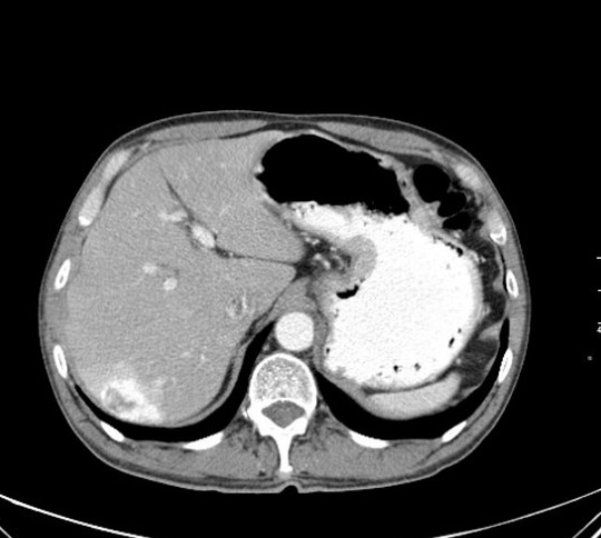
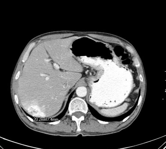


По артериальной фазе контрастирования похоже на гемангиому. Остальные фазы, к сожалению, не представлены.
Добрый админ
Вы абсолютно правы! Это классическая гемангиома печени. Остальные фазы не были опубликованы намеренно. По последним данным; для точной диагностики гемангиом печени, вполне достаточно исследования в артериальную фазу; Вы спросите почему? На эту тему я как раз и предлагаю поговорить.
Let me see...
radiographia.ru
Марио я с Вами соглашусь, даже и своего скромного опыта замечал, что заряжая многофазовое исследование гемангиому узнаешь сразу на артериальной, а все отсальные фазы идут без диагностической ценности.
Видите, я же узнал ее по артериальной фазе, значит действительно, наверное достаточно =)
Теперь хотелось бы услышать научные аргументы доктора Марио.
Добрый админ
КТ/МРТ исследования печени требуют чётких и обоснованных протоколов, невозможно все пациентам выполнять ,нативные, ранние/поздние артериальные, портальные и отсроченные фазы. Это трудоёмко, весьма затратно о несёт серьёзную лучевую нагрузку для пациента. Было отмечено, что образования печен с периферическим усилением в артериальную фазу, имеют два типа контрастирования (lesions with peripheral contrast attenuations at arterial phase):
1. contineous peripheral ring-like enhancement: по типу замкнутого кольца: метастазы метастазы и абсцессы (также грибковые поражения, например кандидоз).
2. non-contineous peripheral puddle-like enhancement: по типу не-замкнутого периферического усиления с отдельными "лакунами", иногда с центрипетальными "языками" (centripetal contarst extensions); характерных исключительно для гемангиом.
Вывод: если идёт КТ исследование брюшной полости, и вся желаемая диагностическая информация была получена на поздней артериальной фазе, при обнаружении характерной картины гемангиомы печени, можно остановить исследование и не выполнять отсроченные венозные фазы, экономя деньги, амортизацию аппарата и конечно заботясь о лучевой безопасности пациента.
Let me see...
radiographia.ru
Марио. Вопрос! как быть с мизерными гемангиомами, они в артериальную фазу "вспыхивают" одномоментно, как же тут применить тип контрастирования?. У меня реально существует проблема в диф.диагностики малых образований, толи это гемангиомы, толи гиперваскулярные метастазы (были случаи), причем я ориентировался именно на отсроченные фазы, известно, что гемангиомы выравниваются с плотностью печени, а метастазы нет+ быстро "вымывают" контраст.
Интересный вопрос, в таких случаях (множественные гиперваскулярные образования), отсроченную фазу делать необходимо, исключить метастазы является первейшей задачей. Таk что всё Вы делаете правильно. И заметьте, при мелких множественных гемангиомах, они могут контрастироваться (как Вы правильно заметили "вспыхивать"), полностью, так что говорить о "периферическом типе усиления с лакунами контраста" в таких случаях нельзя, следовательно и правило на сработает.
Let me see...
radiographia.ru
Еще один вопрос. Марио у Вас были ситуации например: на КТ данных за гемангиому нет, а при верификации она есть!?. К тому что все ли гемангиомы, именно ВСЕ и ВСЕГДА имеют патогномоничную картину контрастирования. Бывало что я встречался с такими заключениями как атипичные, гиповаскулярные гемангиомы. Но классификация не подразумевает таких видов гемангиом, и совершенно не понятно на каких признаках выносятся такие заключения.
Атипичные гемангиомы печени:
Вы задали очень сложный вопрос. Мы все прекрасно знаем; что медицина это не математика, всегда будет 5-10% патологии, которая не подходить под классическое описание.
Термин атипическая гемангиома печени; используют в УЗИ диагностике, имеется ввиду такие гемангиомы, которые имеет не-классическую (гиперэхогенную),атипическую презентацию. Как правило они в разной степени гипоэхогенны, особенно в центральной части (могут быть гетерогенными) с гипрэхогенным ободком. Такая презентация объясняется последствиями центрального геморрагического некрозом, формированием рубцов или миксоматозными изменениями. Некоторые авторы заявляют, что процент таких атипичных гемангиом достигает 40% (!) (Moody and Wilson 1993).
В отношении КТ и МРТ; более распространён термин: гемангиомы с атипическим контрастированием; как правило такая атипия объясняется изменениями в окружающей печёночной ткани, например наличие фиброзных изменений за счёт активного вирусного гепатита С: посмотрите эту статью (можно скачать .PDF):
http://www.springerlink.com/content/n11n762475112635/
Masakatsu Tsurusaki1, 3 Contact Information, Ryota Kawasaki1, Masato Yamaguchi1, Koji Sugimoto1, Takumi Fukumoto2, Yonson Ku2 and Kazuro Sugimura1
(1) Department of Radiology, Kobe University Graduate School of Medicine, 7-5-2 Kusunokicho, Chuo-ku, Kobe 650-0017, Japan
(2) Department of Hepato-Biliary and Pancreatic Surgery, Kobe University Graduate School of Medicine, Kobe, Japan
(3) Present address: Department of Diagnostic Radiology, National Cancer Center, 5-1-1 Tsukiji, Chuo-ku, Tokyo 104-0045, Japan
Received: 29 September 2008 Accepted: 10 December 2008 Published online: 3 May 2009
Abstract We report a case of hemangioma with an atypical vascular enhancement pattern. The hemangioma showed peripheral rim enhancement at the arterial phase during dynamic magnetic resonance imaging, and the peripheral enhanced zone was still apparent during the delayed phase, as shown on double-phase computed tomography hepatic arteriography. The rim enhancement pattern of this case, mimicking that of hepatocellular carcinoma, may be due to the surrounding liver parenchymal fibrotic change caused by an active hepatitis C viral infection.
Другие разновидности гемангиом которые могут попадать под термин атипические; включают в себя:
1.Быстро накапливающие гемангиомы (как Вы написали "вспыхивающие").
2.Медленно накапливающие гемангиомы (до 24 часов!), их бывает очень сложно дифференцировать.
3.Кальцинированные гемангиомы: редко встречаются, кальцинация может быть как по периферии, так и в центре (по типу флеболитов), в диагностике таких гемангиом помогает МРТ, мы увидим гиперинтенсивное на Т2W образование с кальцинацией.
4.Гиалинизированная гемангиома: считается что гиалинизирование, это последняя стадия инволюции гемангиомы, патоморфологи описывают выраженные процессы фиброза с облитерацией сосудов. Такие гемангиомы практически невозможно диагностировать при помощи радиологических методов (Вы об этом как раз писали).
5.Кистозные и мультикистозные гемангиомы: очень редкий вариант, мультикистозная гемангиома была описана только единожды (Hihara et al. 1990), отличить от других вариантов кист печени практически невозможно.
6.Гемангиомы с уровнями жидкости: также очень редкий вариант, на УЗИ выглядят гипоэхогенными, уровни выявляются только на КТ и МРТ.
7.Гемангиомы с артерио-венозным шунтированием: хотя артерио-венозное шунтирование связывают с злокачественными образованиями, этот феномен описан и при таких доброкачественных образованиях, как гемангиомы. Данные феномен можно визуализировать на динамическом КТ с усилением или МРТ с контрастированием; наблюдается раннее паренхиматозное контрастирование с наполнением портальной вены. В литературе описано, что > 25% гемангиом могут наблюдаться с таким феноменом, чаще в гемангиомах с быстрым наполнением.
8.Гемангиомы с ретракцией капсулы: описаны при ассоциации гемангиомы с злокачественными опухолями (холангиокарцинома, гемангиоэндотелиома и метастазами); существует всего несколько наблюдений описанных в литературе.
Я постарался более менее ответить на Ваш вопрос; и благодарен Вам, так как сам узнал для себя новую информацию, в процессе подготовки этого поста.
Если Вы владеете английским, я представляю Вашему вниманию две статьи по теме, где наиболее полно раскрыта эта тема.
http://radiographics.rsnajnls.org/cgi/content/full/20/2/379
http://www.ajronline.org/cgi/content/full/180/1/135
What's this?
AJR 2003; 180:135-141
© American Roentgen Ray Society
Pictorial essay
Hepatic Hemangioma: Atypical Appearances on CT, MR Imaging, and Sonography
Hyun-Jung Jang1,2, Tae Kyoung Kim3, Hyo Keun Lim4, Sang Jae Park2, Jung Suk Sim1, Hyae Young Kim1 and Joo-Hyuk Lee1
1 Radiation Medicine Branch, Research Institute, National Cancer Center, 809 Madu 1-dong, Ilsan-gu, Goyang-si, Gyeonggi-do, 411-764, Korea.
2 Center for Liver Cancer, National Cancer Center Hospital, National Cancer Center, Gyeonggi-do, 411-764, Korea.
3 Department of Diagnostic Radiology, Asan Medical Center, University of Ulsan College of Medicine, 388-1, Poongnap-dong, Songpa-gu, Seoul, 138-736, Korea.
4 Department of Radiology and Gastrointestinal Center, Sungkyunkwan University School of Medicine, Samsung Medical Center, 50 Ilwon-dong, Kangnam-gu, Seoul, 135-710, Korea.
Received April 25, 2002; accepted after revision July 2, 2002.
Address correspondence to H.-J. Jang.
Introduction
Hemangioma is the most common benign tumor of the liver. The classic diagnostic findings for hemangioma are as follows [1]: on unenhanced CT, hypoattenuation similar to that of vessels; on dynamic contrast-enhanced CT or MR imaging, peripheral globular enhancement and a centripetal fill-in pattern with the attenuation of enhancing areas identical to that of the aorta and blood pool; on T2- and heavily T2-weighted MR imaging, hyperintensity similar to that of cerebrospinal fluid; on sonography, homogeneous hyperechogenicity or hypo- or isoechogenicity with a hyperechoic rim; and on delayed phases of 99mTc RBC scanning, a defect in the early phases that shows prolonged and persistent filling-in. Because of advances in imaging technology, hemangiomas are being detected more frequently. We have encountered various atypical forms that may be difficult to recognize as hemangiomas on cross-sectional imaging. In this pictorial essay, we illustrate the varied appearances of hemangiomas that do not meet conventional criteria on various current imaging techniques and provide possible explanations for their atypical appearances.
Small Hypoattenuating Hemangioma
Small hemangiomas are detected more frequently with helical CT, whereas they are easily overlooked on conventional CT because they tend to be isoattenuating on late-phase images [2]. Due to earlier scanning, slowly enhancing hemangiomas have more chance to show persistent hypoattenuation, the incidence being up to 8-16% [2, 3]. In daily practice, the incidence of this form of hemangioma is even greater than previously reported, especially for small hemangiomas that may not show the classic rapid-fill-in pattern [2]. The reason for this reported lower incidence is likely that the atypical appearance of this type of hemangioma may have misled researchers into precluding the possibility of hemangioma in the first place.
Small hypoattenuating hemangiomas are particularly problematic in patients with underlying malignancy. If present, the "bright-dot" sign—tiny enhancing dots in the hemangioma that do not progress to the classic globular enhancement because of the small size of the lesion and the propensity for very slow fill-in—is helpful in diagnosing this type of hemangioma [2] (Fig. 1A,1B,1C,1D). However, a number of hemangiomas have no discernible enhancement (Fig. 2A). One pathologic correlative study suggested that hemangiomas with a slow fill-in pattern have relatively large vascular spaces and that those with rapid enhancement have small vascular spaces and a large interstitium [4]. Such a tendency has no relationship to the size of the tumor [4]. Therefore, hemangioma should be included in the differential diagnoses of small hypoattenuating lesions as well as hypervascular lesions. Contrast-enhanced gray-scale harmonic sonography, which has the capability of real-time dynamic assessment, could be of help in characterizing such a small hypoattenuating hemangioma seen on routine single-phase helical CT (Figs. 2B and 2C).
View larger version (125K):
[in this window]
[in a new window]
[as a PowerPoint slide]
View larger version (135K):
[in this window]
[in a new window]
[as a PowerPoint slide]
View larger version (136K):
[in this window]
[in a new window]
[as a PowerPoint slide]
View larger version (124K):
[in this window]
[in a new window]
[as a PowerPoint slide]
View larger version (101K):
[in this window]
[in a new window]
[as a PowerPoint slide]
View larger version (181K):
[in this window]
[in a new window]
[as a PowerPoint slide]
View larger version (183K):
[in this window]
[in a new window]
[as a PowerPoint slide]
Atypical Signal on T2-Weighted MR Imaging
A long T2 relaxation time has been attributed to the presence of slowly flowing blood in the vascular spaces of the tumor, and this bright T2 signal on MR imaging is one of the most reliable findings in diagnosing hemangioma [5]. It has been reported that a threshold of 112 msec of T2 relaxation time results in 92% accuracy, 96% sensitivity, and 87% specificity for differentiating hemangiomas from metastases [5]. In small hemangiomas, marked hyperintensity on T2-weighted images is a particularly important finding because the pathognomonic nodular enhancement is frequently not present [3]. Rarely, hemangiomas with rapid enhancement (Fig. 3A,3B) or with unusual abnormalities (Fig. 4A) may show T2 signal intensity that is not as bright as cerebrospinal fluid on MR imaging and may cause confusion. The signal intensity characteristics are known to be related to the relative composition of vascular spaces and connective tissue in the lesion and to the presence of thrombosis, calcification, hemorrhage, or fibrosis [5].
View larger version (162K):
[in this window]
[in a new window]
[as a PowerPoint slide]
View larger version (145K):
[in this window]
[in a new window]
[as a PowerPoint slide]
View larger version (125K):
[in this window]
[in a new window]
[as a PowerPoint slide]
Attenuation Relative to Vascular Pool
Because enhancing areas in hemangioma consist of vascular spaces directly supplied by arteries, the attenuation of such areas is theoretically identical to that of the aorta on hepatic arterial phase and that of blood pool during later phase imaging. Such a characteristic is helpful in differentiating hemangiomas from other tumors, but occasionally this finding makes it difficult to distinguish hemangiomas from vessels on CT (Fig. 5). On the other hand, not rarely for hemangiomas in general, and more commonly in small hemangiomas, the enhancing areas show lower attenuation than that of the aorta or portal or hepatic veins on multiphase helical CT [3] (Figs. 4B and 6).
View larger version (134K):
[in this window]
[in a new window]
[as a PowerPoint slide]
View larger version (155K):
[in this window]
[in a new window]
[as a PowerPoint slide]
View larger version (157K):
[in this window]
[in a new window]
[as a PowerPoint slide]
Hemangioma Versus Hypervascular Malignancy
Differentiation of hemangiomas from other hypervascular tumors can be a challenge because some hypervascular tumors can mimic peripheral globular enhancement (Fig. 7A,7B,7C), and not all hemangiomas show such a characteristic pattern [3, 5]. Neuroendocrine tumors or metastases from breast or colon cancer may show strong T2 hyperintensity [5], and prolonged contrast-enhancement may be seen in certain hypervascular malignancies [3]. It is helpful to know that hemangiomas can remain unenhanced, but once the areas enhance they do not diminish. Interpretation based on the combination of two or more imaging characteristics is required.
View larger version (158K):
[in this window]
[in a new window]
[as a PowerPoint slide]
View larger version (149K):
[in this window]
[in a new window]
[as a PowerPoint slide]
View larger version (106K):
[in this window]
[in a new window]
[as a PowerPoint slide]
Hemangioma with Arterioportal Shunt
An arterioportal shunt associated with a hepatic tumor is generally recognized to be most characteristic of malignant tumors. However, an arterioportal shunt sometimes is seen in hepatic hemangiomas on multiphase helical CT [6] (Fig. 8A,8B). Similar temporal peritumoral enhancement can be seen on dynamic MR images [7] (Fig. 9A,9B). These tumors tend to show rapid enhancement [6, 7]. One possible explanation for this finding is that a rapidly enhancing small hemangioma has hyperdynamic status with large arterial inflow, rapid tumoral enhancement, and consequently, large and rapid outflow, which seems to result in early opacification of the draining portal vein via shunt and peritumoral enhancement [7]. The finding to note is a wedge-shaped or irregularly shaped enhancement adjacent to the hemangioma—with or without early visualized portal branches—during the hepatic arterial phase (Fig. 8A,8B) that becomes isoattenuating or slightly hyperattenuating relative to the normal liver during the portal venous phase [6]. An association with arterioportal shunt does not necessarily imply that the underlying tumor is malignant.
View larger version (138K):
[in this window]
[in a new window]
[as a PowerPoint slide]
View larger version (141K):
[in this window]
[in a new window]
[as a PowerPoint slide]
View larger version (138K):
[in this window]
[in a new window]
[as a PowerPoint slide]
View larger version (135K):
[in this window]
[in a new window]
[as a PowerPoint slide]
Hemangioma in Fatty Liver
Severe fatty liver may alter the apparent enhancement pattern of focal hepatic lesions. Even hypovascular tumors such as metastases can show relatively high attenuation on CT and may mimic hemangiomas with a persistent enhancement pattern (Fig. 10A,10B). In severe fatty liver, the attenuation of hemangioma may reverse to even hyperattenuation, although not greater than that of vessels, on unenhanced CT. Hemangiomas may also be accompanied by a focal spared zone as seen in malignant tumors in fatty liver. On sonography, this finding could create confusion with the hypoechoic halo seen in malignant tumors (Fig. 11A,11B,11C), contrary to hemangiomas' usual hyperechogenicity or hyperechoic rim. This unusual finding often makes subsequent CT or MR imaging necessary. Hemangiomas in fatty liver could produce a peculiar halo on CT or MR imaging as well, but in most cases, accurate diagnosis can be made without difficulty because of the characteristic dynamic enhancement pattern of hemangiomas.
View larger version (135K):
[in this window]
[in a new window]
[as a PowerPoint slide]
View larger version (128K):
[in this window]
[in a new window]
[as a PowerPoint slide]
View larger version (166K):
[in this window]
[in a new window]
[as a PowerPoint slide]
View larger version (128K):
[in this window]
[in a new window]
[as a PowerPoint slide]
View larger version (133K):
[in this window]
[in a new window]
[as a PowerPoint slide]
Hemangioma in Liver Cirrhosis
With progressive cirrhosis, hemangiomas are likely to decrease in size and become more fibrotic (Fig. 12A,12B,12C) and difficult to diagnose radiologically and pathologically [8]. Conversely, hepatocellular carcinoma and dysplastic nodules often mimic hemangioma on sonography (Fig. 13A,13B) because of hyperechogenicity resulting from factors such as necrosis, fibrosis, fatty change, or sinusoid dilatation. In cirrhosis, any hyperechoic nodule should be considered a probable hepatocellular carcinoma until proven otherwise.
View larger version (161K):
[in this window]
[in a new window]
[as a PowerPoint slide]
View larger version (178K):
[in this window]
[in a new window]
[as a PowerPoint slide]
View larger version (162K):
[in this window]
[in a new window]
[as a PowerPoint slide]
View larger version (143K):
[in this window]
[in a new window]
[as a PowerPoint slide]
View larger version (179K):
[in this window]
[in a new window]
[as a PowerPoint slide]
Conclusion
A subset of hepatic hemangiomas does not show the classic findings on CT, MR imaging, and sonography that are well known to radiologists. Radiologists should be aware that some hepatic hemangiomas may have atypical features on cross-sectional imaging that correlate with their varied hemodynamic and pathologic findings.
References
Gore RM, Levine MS. Textbook of gastrointestinal radiology, 2nd ed. Philadelphia: Saunders, 2000: 1487-1497 Jang H-J, Choi BI, Kim TK, et al. Atypical small hemangiomas of the liver: "bright dot" sign at two-phase spiral CT. Radiology 1998;208:543 -548[Abstract/Free Full Text] Kim T, Federle MP, Baron RL, Peterson MS, Kawamori Y. Discrimination of small hepatic hemangiomas from hypervascular malignant tumors smaller than 3 cm with three-phase helical CT. Radiology 2001;219:699 -706[Abstract/Free Full Text] Yamashita Y, Ogata I, Urata J, Takahashi M. Cavernous hemangioma of the liver: pathologic correlation with dynamic CT findings. Radiology 1997;203:121 -125[Abstract/Free Full Text] Bennett GL, Petersein A, Mayo-Smith WW, Hahn PF, Schima W, Saini S. Addition of gadolinium chelates to heavily T2-weighted MR imaging: limited role in differentiating hepatic hemangiomas from metastases. AJR 2000;174:477 -485[Abstract/Free Full Text] Kim KW, Kim TK, Han JK, Kim AY, Lee HJ, Choi BI. Hepatic hemangiomas with arterioportal shunt: findings at two-phase CT. Radiology 2001;219:707 -711[Abstract/Free Full Text] Jeong M-G, Yu J-S, Kim KW. Hepatic cavernous hemangioma: temporal peritumoral enhancement during multiphase dynamic MR imaging. Radiology 2000;216:692 -697[Abstract/Free Full Text] Brancatelli G, Federle MP, Blachar A, Grazioli L. Hemangioma in the cirrhotic liver: diagnosis and natural history. Radiology 2001;219:69 -74[Abstract/Free Full Text]
This article has been cited by other articles:
S. H. Kim, J. M. Lee, J. Y. Lee, J. K. Han, S. K. An, C. J. Han, K. H. Lee, S. S. Hwang, and B. I. Choi
Value of Contrast-Enhanced Sonography for the Characterization of Focal Hepatic Lesions in Patients with Diffuse Liver Disease: Receiver Operating Characteristic Analysis
Am. J. Roentgenol., April 1, 2005; 184(4): 1077 - 1084.
[Abstract] [Full Text] [PDF]
J. H. Byun, T. K. Kim, C. W. Lee, J. K. Lee, A. Y. Kim, P. N. Kim, H. K. Ha, and M.-G. Lee
Arterioportal Shunt: Prevalence in Small Hemangiomas versus That in Hepatocellular Carcinomas 3 cm or Smaller at Two-Phase Helical CT
Radiology, August 1, 2004; 232(2): 354 - 360.
[Abstract] [Full Text] [PDF]
J. C. Vilanova, J. Barcelo, J. G. Smirniotopoulos, R. Perez-Andres, M. Villalon, J. Miro, F. Martin, J. Capellades, and P. R. Ros
Hemangioma from Head to Toe: MR Imaging with Pathologic Correlation
RadioGraphics, March 1, 2004; 24(2): 367 - 385.
[Abstract] [Full Text] [PDF]
Figures Only
What's this?
Let me see...
radiographia.ru
Марио очень благодарен за Ваш объемный ответ, чувствую себя виноватым перед Вами, что заставил Вас уделить столько времени на подготовку поста.
Вы не правы! Когда отвечаешь на такие вопросы, сам учишься, это я Вам благодарен за то что заставили в очередной раз перелопатить современные источники; я сам узнал для себя новую или подзабытую информацию. Надеюсь что представленная информация пригодится в Вашей работе.
С Уважением.
Let me see...
radiographia.ru