Иллюстрации. ГМ. Нейровизуализация для студентов.
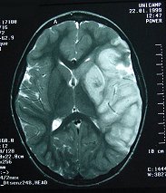
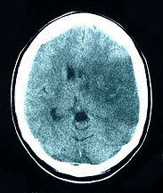
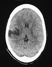
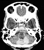
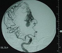
ISCHEMIC CEREBRAL INFARCTIONS
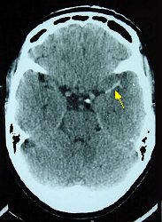
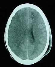
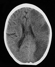
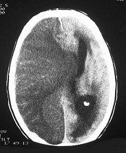
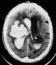
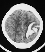
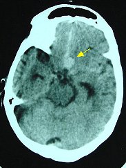
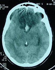
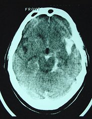
Recent ischemic infarct due to thrombosis of the left middle cerebral artery (less than 12 hours) Recent ischemic infarct due to lesion of internal carotid A. in skull fracture Ischemic infarct of middle cerebral A. due to meningitisRecent ischemic hemispheric infarct with massive edema and subfalcine
herniation
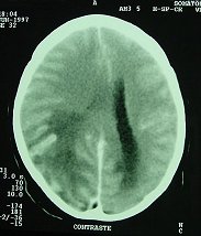
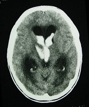
Acute infarct of middle cerebral A due to traumatic thrombosis of left internal carotid A. Recent infarct of part of right middle cerebral A. territoryRecent infarct of part of left middle cerebral A. territorySubacute embolic infarct of temporal branches of middle cerebral A. 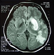
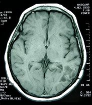
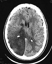
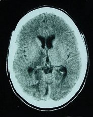
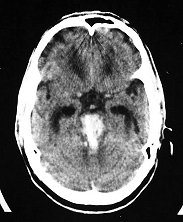
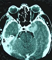
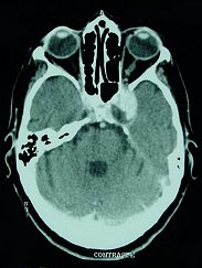
Recent left sided infarct of lenticulostriate A. and cortical branches of middle cerebral A.Infarct of cortical branches of middle cerebral A. Acute phase and after 4 monthsRecent ischemic infarct of right posterior cerebral A. Post-traumatic ischemic infarct of both posterior cerebral Aa. 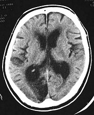
Old sequelar infarct of posterior cerebral A. Acute bilateral infarction of anterior cerebral Aa. due to vasospasm after aneurysm rupture.
..
STATUS LACUNARIS
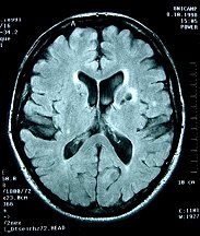
Status lacunaris of basal ganglia
..
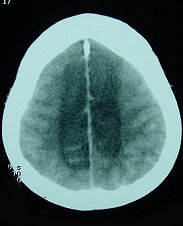
HEMORRHAGIC CEREBRAL INFARCTIONS
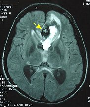
Frontal hemorrhagic infarct due to superior sagittal sinus thrombosisHemorrhagic infarct of perisylvian regionHemorrhagic infarct of posterior cerebral A. (calcarine infarction) secondary to chronic subdural hematoma and uncal herniation Hemorrhagic infarct of both anterior cerebral Aa. after rupture of aneurysm of anterior communicating A. and vasospasm.
..
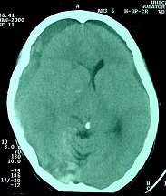
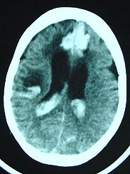
CEREBRAL HEMORRHAGE
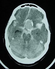
Classical hypertensive capsulolenticular hemorrhage in basal ganglia Hypertensive hemorrhage in basal ganglia and thalamus with ventricular ruptureThalamic hypertensive hemorrhage with ventricular ruptureBrain stem hypertensive hemorrhage with ventricular rupture 
Brain stem hypertensive hemorrhageCerebral hemorrhages in acute leukemia
..
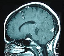
ANEURYSMS AND SUBARACHNOID HEMORRHAGE
Partly thrombosed aneurysm of left intracavernous internal carotid A. Intracavernous aneurysm of internal carotid A. with parietal thrombus Giant aneurysm of internal carotid A. with subarachnoid and ventricular hemorrhage Aneurysm of anterior communicating A. with subarachnoid and ventricular hemorrhage
Aneurysm of anterior communicating A. with vasospasm. Bilateral infarct of anterior cerebral Aa.Saccular aneurysm of anterior cerebral A branchAneurysm of internal carotid A. bifurcation with subarachnoid hemorrhage Basal subarachnoid hemorrhage with ventricular rupture
..
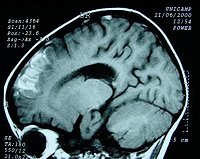
CAVERNOMAS
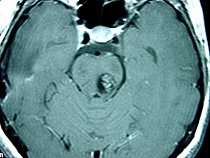
Pontine cavernoma with medullary infarctionCavernoma of middle frontal gyrus
..
ARTERIOVENOUS MALFORMATIONS (AVMs)
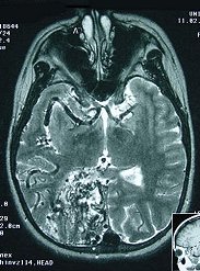
Massive arteriovenous malformation of right cerebral hemisphere Midline parietal arteriovenous malformation
..
..
..
..
..
..
..
..
..
..
..
..
..
..
Dept of Anatomy
IB - UNICAMP
Dept of Pathology
FCM - UNICAMP
Dept of Anatomy
IB - UNICAMP
Dept of Pathology
FCM - UNICAMP
Collaboration
Dept of Pathology
FCM - UNICAMP
Dept of Anatomy, Cell Biology, Physiology and Biophysics, IB - UNICAMP
МР-ангиография сонных артерий и позвоночных
http://www.info-radiologie.ch/carotide-russe.php
Круг Уиллиса (MRA TOF MIP)
http://www.info-radiologie.ch/willis-russe.php
Венозные синусы мозга (МРА)
http://www.info-radiologie.ch/sinus-veineux-russe.php
МРТ гипофиза и области турецкого седла
http://www.info-radiologie.ch/hypophyse-russe.php
Сканирование мозга
http://www.info-radiologie.ch/atlas-tdm-cerveau-ru.php
Придаточные пазухи носа
http://www.info-radiologie.ch/scanner-sinus-ru.php
Нейровизуализация эпилепсии в педиатрии.
http://pubs.rsna.org/doi/full/10.1148/rg.284075114