Астроцитомы. Глиобластома. Глиосаркома. Глиоматоз. Аденомы гипофиза. Опухоли пинеальной области (герминома, тератома). Опухоли шишковидной железы. Параганглиома.
Астроцитома
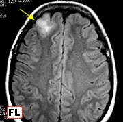
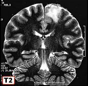
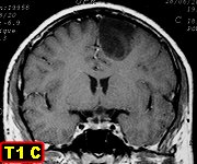
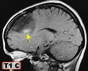 F. 47 yr. Small low grade frontal astrocytoma (incidental MRI finding).M. 35 yr. Small low grade frontal astrocytoma in premotor area M. 33 yr. Frontal mesial low grade diffuse astrocytoma F. 30 yr. Low grade frontal astrocytoma with early anaplasia (foci of contrast enhancement)
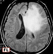
 M. 44 yr. Poorly circumscribed low grade frontal astrocytomaM. 36 yr. Recurrent frontobasal low grade astrocytoma, 6 yr followup M. 7 yr. Low grade solid-cystic astrocytoma of hemispheric white matterM. 43 yr. Low grade diffuse astrocytoma of left hemisphere infiltrating from temporal lobe through thalamus
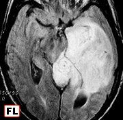
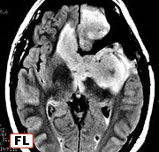
 M. 43 yr. Diffuse temporal astrocytoma, transtentorial herniation and midbrain compression M. 52 yr. Diffuse temporal astrocytoma infiltrating brainstemM. 39 yr. Diffuse low grade temporal astrocytoma with anaplastic recurrence after 4 yearsM. 32 yr. Anaplastic solid-cystic
frontal gemistocytic
astrocytoma (WHO grade III) 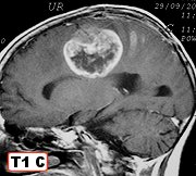
 F. 10 yr. Anaplastic astrocytoma of centrum semiovaleM. 53 yr. Paramedian anaplastic astrocytoma with contralateral extension via corpus callosumM. 68 yr. Anaplastic astrocytoma of corpus callosum infiltrating from hippocampus to hypothalamus via fornixF. 54 yr. Low grade astrocytoma of insular region
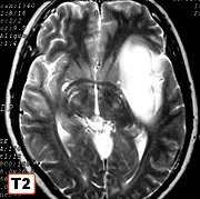
 M. 34 yr. Low grade astrocytoma of insular regionM. 38 yr. Low grade astrocytoma of insular regionF. 40 yr. Low grade astrocytoma of insular region extending into frontal and temporal lobesM. 52 yr. Low grade astrocytoma of thalamus with secondary infiltration of hippocampus and insula
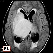
 M. 5 yr. Diffuse low grade astrocytoma of thalamus and basal gangliaF. 51 yr. Low grade astrocytoma of left thalamus extending into centrum semiovale, corpus callosum and right hippocampusF. 43 yr. Bilateral thalamic astrocytomaF. 11 yr. Diffuse astrocytoma of right thalamus with anaplastic foci
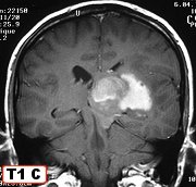
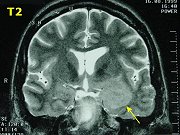
 M. 37 yr. Anaplastic thalamic astrocytomaF. 46 yr. Low grade periaqueductal astrocytoma M. 21 yr. Low grade periaqueductal astrocytoma M. 8 yr. Anaplastic astrocytoma of pons
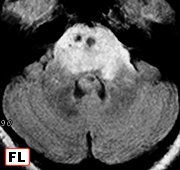
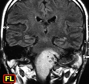
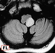
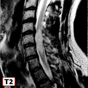 F. 41 yr. Low grade astrocytoma of pons and medullaM. 36 yr. Diffuse astrocytoma of brain stemF. 11 yr. Exophytic anaplastic astrocytoma of lateral medulla oblongataM. 32yr. Low grade astrocytoma of cervical spinal cord
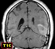
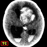
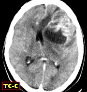
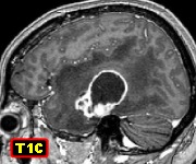
Глиобластома
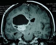
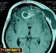
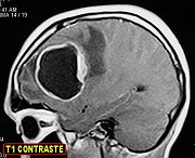
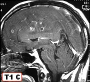
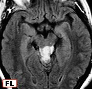
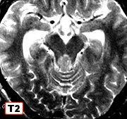
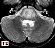
 Glioblastoma multiforme with unusual presentation and rapid courseBilateral frontal glioblastoma multiforme Left frontal giant cell glioblastoma Glioblastoma multiforme of pontine tegmentum
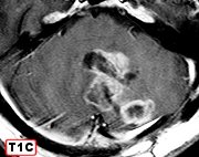
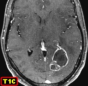
 Glioblastoma multiforme of cerebellumTemporoparietal glioblastoma multiforme F. 59 yr. Left sided temporal glioblastoma multiforme Same case 9 months after surgery and radiotherapy - pseudotumoral radiation necrosis
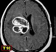
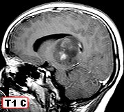
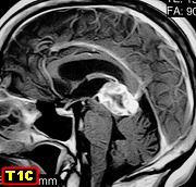 F. 27 yr. Giant cell glioblastomaF. 5 months. Glioblastoma multiforme of left cerebral hemisphere (presumably congenital)F. 58 yr. Giant cell glioblastoma with a component of conventional glioblastoma multiformeF. 64 yr. Glioblastoma multiforme of pineal
Глиосаркома
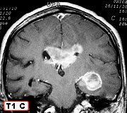
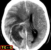
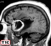
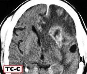
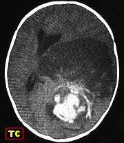
 M. 56 yr. Left temporal gliosarcoma. TextM. 4 d. Congenital gliosarcoma at foramen of Monro, hydrocephalus, spontaneous ventricular bleedingM. 49 yr. Gliosarcoma of left frontal lobeF. 48 yr. Right temporal gliosarcoma. Original MRI
  Same, recurrence 4 months after surgery
GRANULAR CELL ASTROCYTOMA OF BRAIN
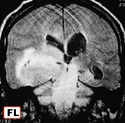
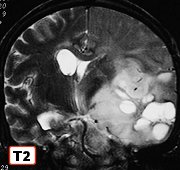
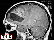
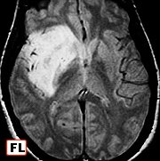
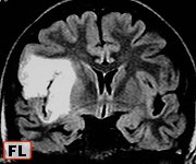
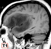
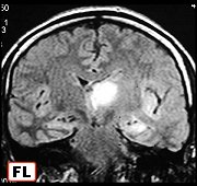
GLIOMATOSIS CEREBRI
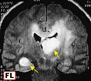
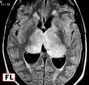
oligoastrocytoma.
Early CT, 6/2004
MRI 4/2006
MRI 6/2006
PRIMARY EXTRACEREBRAL MENINGEAL GLIOMA
Пилоцитарная астроцитома
Зрительный нерв. Пилоцитарная астроцитома
Пилоцитарная астроцитома
with abundant vascular proliferation
pilocytic astrocytoma
Плеоморфная ксантоастроцитома
xanthoastrocytoma-
ganglioglioma-
ependymoma
xanthoastrocytoma
TUBEROUS SCLEROSIS & SUBEPENDYMAL GIANT CELL ASTROCYTOMA
sclerosis of diploe
Аденомы гипофиза
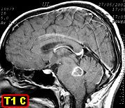
Опухоли пинеальной области (герминома, тератома)
Опухоли шишковидной железы
Параганглиома
Глиобластома
http://radiopaedia.org/images/3062942
http://radiopaedia.org/images/3062916
http://radiopaedia.org/images/3062897
http://radiopaedia.org/images/3062797
http://radiopaedia.org/images/3062751
http://radiopaedia.org/images/3062691
http://radiopaedia.org/images/3062632
Необычные проявления первичной глиобластомы мозга
http://www.surgicalneurologyint.com/article.asp?issn=2152-7806;year=2010;volume=1;issue=1;spage=87;epage=87;aulast=Sanli
Глиобластома
http://www.ajnr.org/content/30/4/808.full
http://www.pediatricneurosciences.com/article.asp?issn=1817-1745;year=20...
http://scielo.isciii.es/scielo.php?pid=S1130-14732009000600004&script=sc...
Первичная глиобластома мозга
http://surgicalneurologyint.com/article.asp?issn=2152-7806;year=2010;vol...
Низкодифференцированные астроцитомы
http://radiopaedia.org/cases/low-grade-astrocytoma
Анапластическая астроцитома
http://radiopaedia.org/cases/anaplastic-astrocytoma
Глиосаркома
http://jnnp.bmj.com/content/74/3/364.full
http://www.scielo.br/scielo.php?pid=S0004-282X2004000400008&script=sci_arttext
http://www.aboutcancer.com/gliosarcoma.htm
http://radiopaedia.org/cases/gliosarcoma-1
http://www.ruralneuropractice.com/article.asp?issn=0976-3147;year=2012;volume=3;issue=1;spage=60;epage=64;aulast=Balasubramaniam
http://radiopaedia.org/images/19627
http://www.healio.com/hematology-oncology/sarcoma/news/print/hematology-oncology/%7B4bcdbc79-ab71-4c41-b87b-e14eb2b6140f%7D/intracranial-hemorrhage-as-the-initial-presentation-of-gliosarcoma-in-ayoung-woman
http://radiopaedia.org/images/835355
http://www.scielo.org.ar/scielo.php?pid=S1850-15322009000200006&script=sci_arttext
http://www.sciencedirect.com/science/article/pii/S1507136710000726
http://radiopaedia.org/cases/gliosarcoma
Первичная глиосаркома
http://www.ncbi.nlm.nih.gov/pmc/articles/PMC2808523/
Вторичная глиосаркома
http://www.najms.org/article.asp?issn=1947-2714;year=2011;volume=3;issue=11;spage=527;epage=530;aulast=Andaloussi-Saghir
http://www.sciencedirect.com/science/article/pii/S0303846712003368
Глиосаркома таламуса у ребенка
http://www.neurologyindia.com/article.asp?issn=0028-3886;year=2012;volume=60;issue=6;spage=674;epage=676;aulast=Neelima
Глиосаркома у ребёнка
http://www.scielo.br/scielo.php?pid=S0004-282X2008000100022&script=sci_arttext
Опухоли шишковидной железы
http://www.ajronline.org/content/186/3_Supplement/S233.full
Шишковидная область
http://www.neurologyindia.com/article.asp?issn=0028-3886;year=2010;volume=58;issue=6;spage=928;epage=932;aulast=Vaghela
Хордома шишковидной железы (эктопическая)
http://surgicalneurologyint.com/article.asp?issn=2152-7806;year=2011;vol...