La clasificación del Ictus, recogidaenTheOxfordshireCommunityStroke Project, es fácil de comprender pero, no tan sencilla de ejecutar cuando el diagnóstico de un ictus se basa exclusivamente en una exploración clínica. Los hallazgos que proporciona la Tomografía Computarizada corroboran o complementan la sospecha clínica, como podemos apreciar en el siguiente ejemplo (Figuras 1 a 10). Esta mujer de 64 años había sido encontrada en su casa inconsciente. Al llegar a Urgencias se solicitó una TC craneoencefálica con la sospecha clínica de TACI derecho. ¿Era correcto el diagnóstico de presunción?
(The findingsthat provides acomputed tomographyexaminationcorroborateor complementthe clinical suspicion ofacute ictus, as seen inthe following images. Figures 1 to 10)
FIGURA 1) En esta imagen se aprecia un área hipodensa que representa un infarto isquémico subagudo (12 horas). La arteria cerebral media derecha aparece trombosada, destacando como una banda densa sobre el parénquima cerebral infartado.
FIGURA 2) Observando con detalle se puede apreciar que el área hipodensa de tejido infartado afecta al territorio de las arterias cerebral media y cerebral anterior derechas. Por tanto se ha producido un TACI derecho. Pero el área hipodensa se extiende tambien paralela a la superficie izquierda de la hoz del cerebro, territorio de la arteria cerebral izquierda. Por tanto hay que añadir también un PACI izquierdo.
(Looking carefully we can observethat thethe infarctedtissueaffects the territoryof themiddle cerebralarteryand the right anterior cerebral artery.Thusthere has been arightTACI. Buta hypodensebandalso extendsparallel to theleft surface of thefalx cerebri, corresponding to the left anterior cerebralartery territory.Thereforewe must addalso aleftPACI)
FIGURA 3) Representación figurada de los respectivos territorios arteriales. El edema vasogénico que produce el infarto ocurrido en el territorio de la arteria cerebral media derecha comprime a las arterias cerebrales anteriores y, posiblemente, ha provocado la oclusión de ambas. Sólo la arteria cerebral posterior derecha permanece intacta.
(Pictorial representationof thearterials territories. Vasogenicedemaproducedby the infarctionoccurred intheright middle cerebralartery territory, compressesthe anterior cerebral arteriesand possiblycaused theocclusionof both.Only the right posterior cerebralarteryremains intact)
FIGURA 4) Las líneas amarillas delimitan los territorios infartados: TACI derecho y PACI izquierdo. Tanta precisión es dificil de obtener en la exploración clínica de una persona inconsciente.
(The yellow linesdelineatetheinfarcted regions:Right TACIand left PACI.Suchaccuracy isdifficult to obtaininthe clinical examinationofan unconscious person)
FIGURA 5) La línea amarilla delimita el territorio de la arteria cerebral posterior derecha que no se ha lesionado porque depende de la circulación posterior.
(The yellow linedelineatesthe territory of theright posterior cerebralartery. It wasnotinjuredbecause it depends onthe posterior circulation.)
FIGURA 6) Hipodensidad del cerebro infartado en un corte más cefálico. TACI derecha y PACI izquierda.
(The infarctedbrainin a morecephalicimage.Right TACI and left PACI).
FIGURA 7) Hipodensidad del cerebro infartado en un corte más cefálico. TACI derecha y PACI izquierda.
(The infarctedbrainin a morecephalicimage.Right TACI and left PACI).
FIGURA 8) En esta imagen se aprecia también un área hipodensa en el territorio de la arteria cerebral posterior izquierda, secuela de un antiguo infarto. POCI izquierdo.
(Thisimage showsa hypodense areainthe territory of theleftposterior cerebralartery, sequel toan oldinfarction. A left POCI)
FIGURA 9) En esta imagen se aprecia perfectamente el territorio irrigado por cada arteria CMD, CAD y CAIzq.
(Thisimage delineatesperfectlythe territorysupplied byeach arteryMCA, Right ACA andleft ACA)
FIGURA 10) El área infartada se extiende hasta la convexidad de ambos hemisferios cerebrales.
(Theinfarcted areaextends upthe convexityof both cerebral hemispheres)
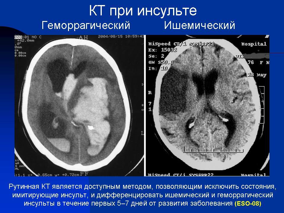
El "ictus" es una urgencia grave que debe ser diagnósticada con prontitud y eficacia, porque en el caso de una accidente isquémico las células del sistema nervioso pueden resistir sin aporte de oxígeno de tres a cinco horas. La mayoría de los hospitales disponen de las dos herramientas de diagnóstico más eficaces que existen actualmente para detectar un "ictus". La primera, la Tomografía Computarizada (TC) es la prueba de elección en todos los casos porque es muy rápida (apenas un minuto de duración) y permite detectar las hemorragias intracraneales con gran precisión. En cambio no tiene sensibilidad para descubrir los pequeños infartos isquémicos encefálicos, hasta que han transcurrido más de doce horas. Y para entonces ya es muy tarde para administrar un tratamiento fibrinolítico preventivo.
FIGURA 1) Paciente de 66 años que fue llevado a Urgencias por sus familiares que lo encontraron inconsciente en la cama. La TC mostró una pequeña área hipodensa en la protuberancia apenas perceptible ¿Infarto o Tumor? (Tiempo de Adquisición 30 segundos)
(A 66-year-old patient was takento the emergency roomby his relatives, who found himunconsciousin bed.The CT scan showeda smallhypodense areain the pons. Infarctionorneoplasm?.) (TAD 30 seconds)
La segunda, la Tomografía por Resonancia Magnética (TRM) es la gran alternativa y resulta de gran ayuda porque es capaz de descubrir un foco isquémico en el encéfalo a la media hora de haberse producido los síntomas. La secuencia SE-EPI potenciada en Difusión Isotrópica (DWI) es la más decuada, porque con ella se pueden obtener 25 imágenes axiales craneoencefálicas en menos de tres minutos. Y si el paciente no colabora y se mueve, la mayoría de los aparatos de TRM modernos disponen una aplicación clínica, SE-EPI- PROPELLER, que elimina los artefactos de movimiento y calcula automáticamente las cifras del Coeficiente de Difusión Aparente (CDA).
A stroke, encephalovascular accident (EVA) or cerebrovascular accident (CVA), is apathological disordercharacterized byacutefailure of theblood supply to theencephalon.Thisdisturbancecanbeduetosudden interruption ofblood flow (ischemia), caused bythrombosisorembolism, orvascular rupture, resulting in acutebleeding (haematoma). Astrokeis anemergencythatmust be diagnosedquicklyand accurately, because the cellsof nervous tissuecan not survivemore than threehours withoutoxygenthat givesblood.
The rapiditywith which itestablishesthe diagnosisis vitalin the results.Ischemic eventscan be solved withfibrinolytic therapy, which is most effective whenit isadministeredbefore. We have twodiagnostic modalities, CT andMRI.Computed Tomography isfaster and hasprioritybecause it allowsrule outahemorrhagicstroke. However, it hasa greatinconvenient: it is notcapable of detectingan ischemic areauntil it has transcurredmore than twelve hours. And thenit's toolate to administerpreventive treatment.
In contrast, MRIis very sensitive andcan detectan ischemic strokeon the half hour after the blood flow has been interrupted.With a sequence ofdiffusion-weighted images (DWI)is sufficient to detectthe focusof ischemia in only three minutes.And if the patientdoes not cooperateand moves,we canusetheSE-EPI- PROPELLER, which eliminatesmotion artifacts.
FIGURA 2) Al medir la densidad se observó una diferencia notable entre el lado derecho de la protuberancia (26 UH) y la pequeña lesión (16 UH).(By measuringthe density we found asignificantdifferencebetweenthe right side ofthe "pons"(26HU)andthe small lesion(16HU).
FIGURA 3) Secuencia Fast Espín Eco potenciada en T2. En esta potenciación brillan los liquidos fisiológicos como el líquido cefalorraquídeo y el humor acuoso de los globos oculares pero también el que se acumula en cualquier proceso patológico. En este caso brilla (hiperseñal) una pequeña lesión redondeada que se localiza en la protuberancia (flecha). Podría ser un infarto isquémico. Ahora bien la pregunta que surge a continuación es la siguiente ¿es un proceso agudo o crónico?. Imposible saberlo porque en ambos supuestos brillaría. Por tanto la imagen potenciada en T2 no nos saca de dudas. (Tiempo de Adquisición 3m 20 seg)
(Axial imageobtained with a T2 weighted FastSpinEchosequenceT2. In thispotentiationenhancementis produced in physiologicalfluidsaccumulated in some anatomical spaces, such as cerebrospinal fluid of ventriclesand aqueous humorofthe eyeballs. But also the waterthat accumulates inanydisease process.In this axial T2 image,we can see an hyperintense small round lesionthat is locatedin the pons (arrow). It could be anischemic stroke. But the questionthat arises the nex is this: is itan acuteor chronic stroke?.Impossible to know,because inboth cases it´llshine.So,T2-weightedimagedoes notget us outof doubt).(TAD 3min 20 sec)
FIGURA 4) Secuencia FLAIR- T2. Con la secuencia FLAIR se anula la señal de los líquidos fisiológicos y sólo brilla el agua que se acumula en las lesiones, sean agudas o crónicas. El posible infarto se aprecia mejor, en esta imagen, pero tampoco salimos de dudas.(Tiempo de Adquisición 4m 40 seg). (T2 weightedFLAIRsequence. WithFLAIR sequence thesignalofphysiologicalfluids is annulled(the basal cisterns andthe eyeballsare shown inblackcolor, very strong) and onlythewater that collectson the round lesion shines. The possible ischemicstrokeis seenbetter,but neitherthis sequencetakes us out ofdoubt.) (TAD 4min 40 sec)
FIGURA 5) Secuencia Fast Espín Eco potenciada en Densidad Protónica. Repetimos una tercera adquisición con una secuencia FSE-Dp. Las imágenes obtenidas tampoco nos aclaran nada acerca de la naturaleza de la lesión que aparece hiperintensa por el aumento en la densidad de moléculas de agua y, como es lógico, de protones. (Tiempo de Adquisición 2m 20 seg). (Proton Density enhanced Fastspin-echosequence. We have repeat a thirdacquisition withFSE-Dp sequence. The images obtainednotclear about the natureof the lesion, thatappearshyperintenseby the increase inthe density ofwater molecules and, as expected,protons). (TAD min 20 sec)
FIGURA 6) Secuencia SE-EPI, potenciada en Difusión Isotrópica (DWI). La lesión brilla intensamente lo cual indica, con los antecedentes clínicos, que se trata de un infarto agudo. Duda solucionada. En estos casos las imágenes potenciadas en Difusión son las más sensibles para detectar un área de isquemia encefálica aguda, en la primera hora de haberse producido los síntomas.(Tiempo de Adquisición 40 seg).
(Diffusion-weighted magnetic resonanceimaging (MRI)provides this image. We can see an enhanced, round lesion in the "pons" indicatinganacute infarction.Doubtresolved.In these cases thediffusion-weighted images (DWI)are most sensitivefor detecting anacute brainischemic areain the firsthour aftersymptomsappeared).(TAD 40 sec).


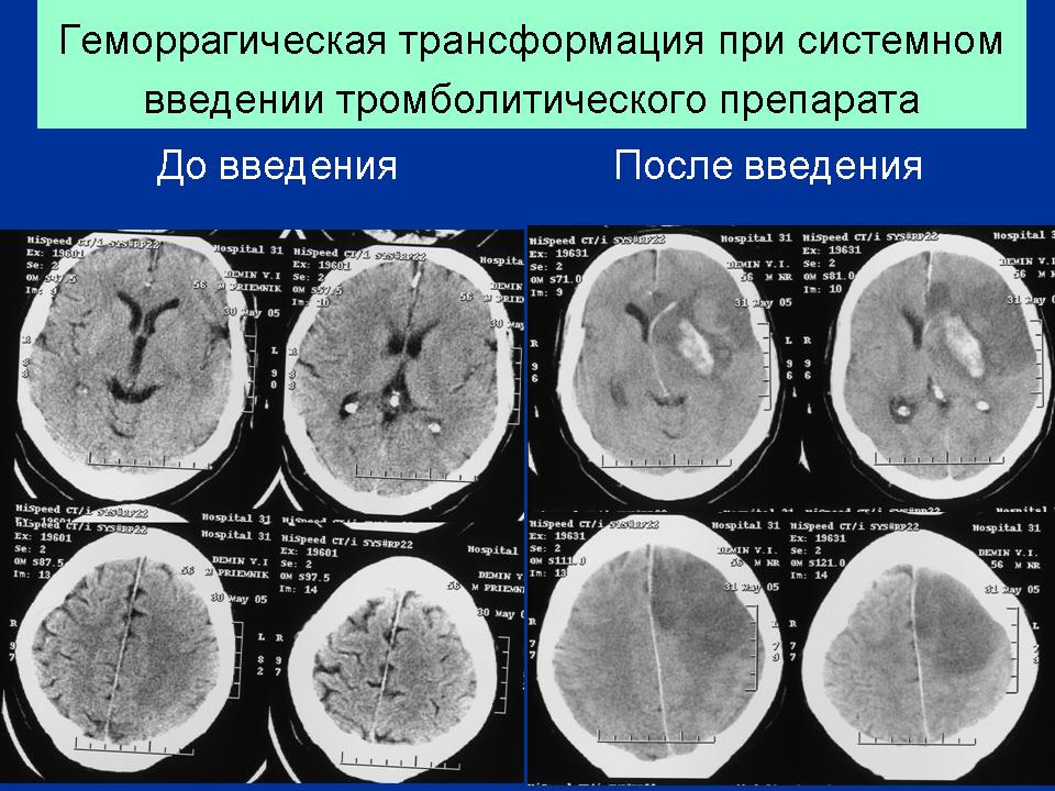
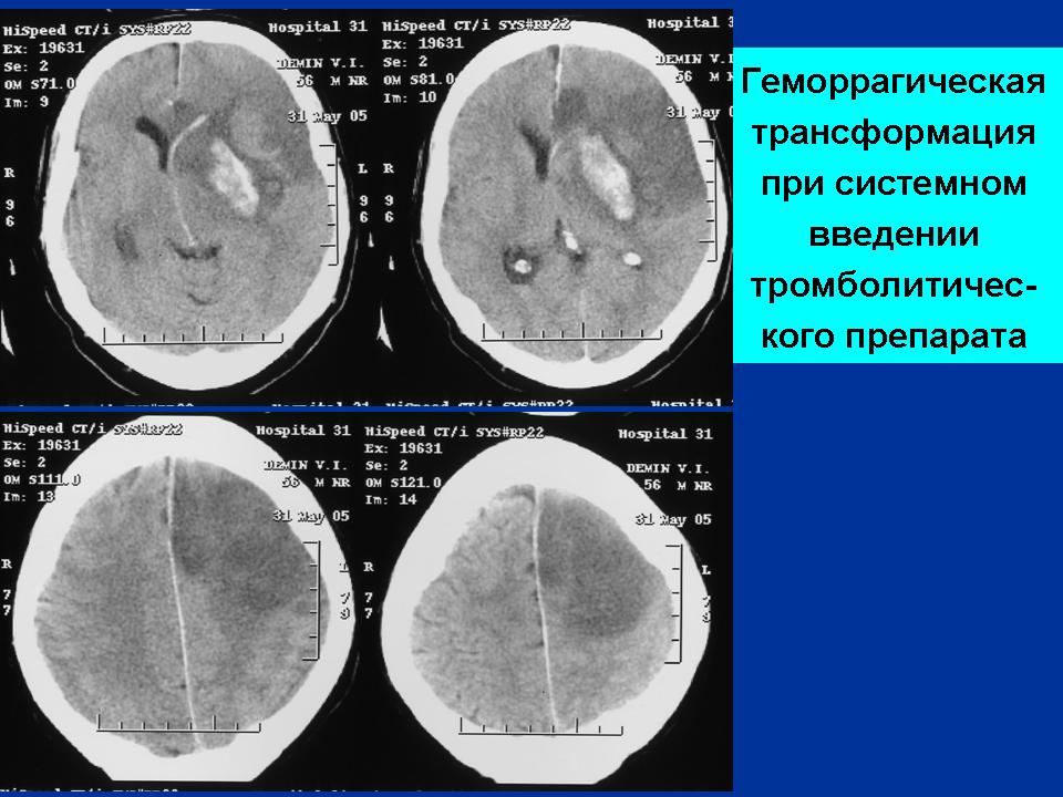
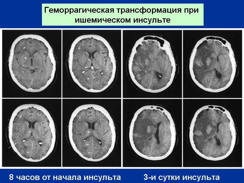
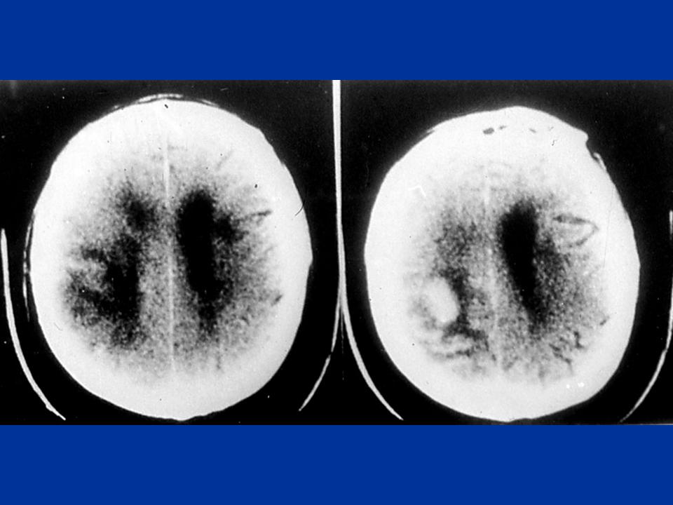
Наблюдение Магонова Е.П.
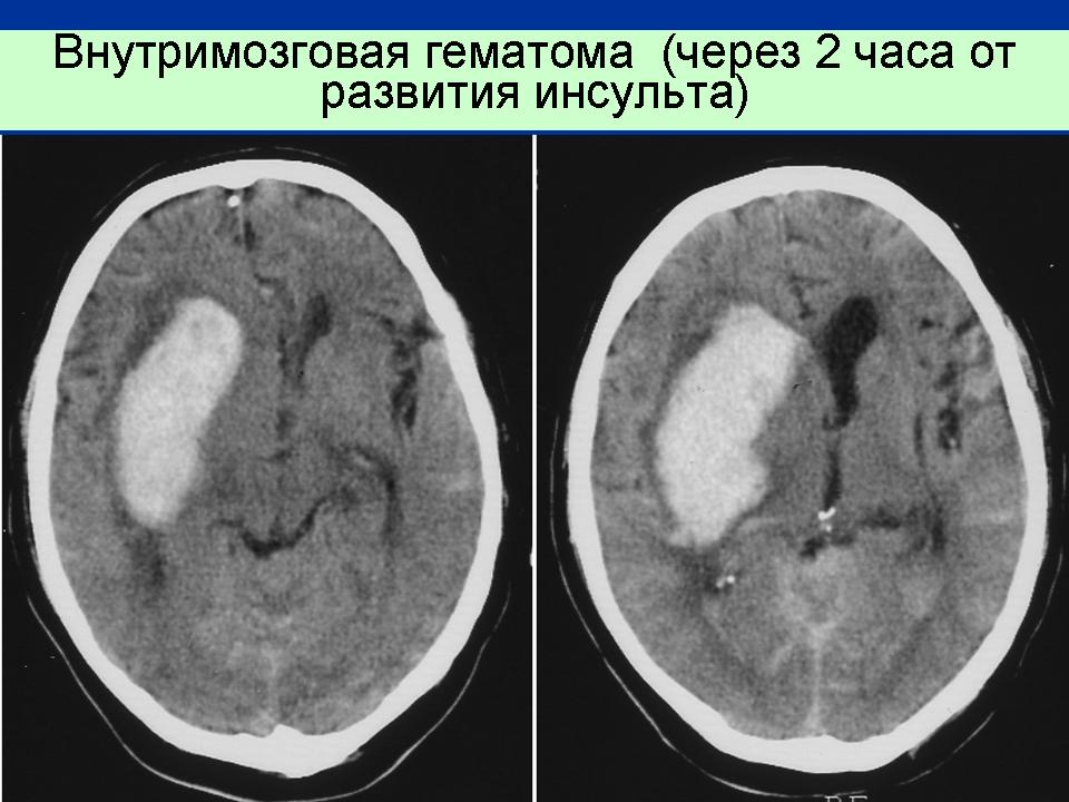
Инфаркт головного мозга (ишемический)
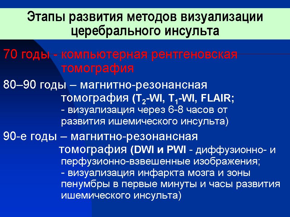
Классификация инсультов
La segunda, la Tomografía por Resonancia Magnética (TRM) es la gran alternativa y resulta de gran ayuda porque es capaz de descubrir un foco isquémico en el encéfalo a la media hora de haberse producido los síntomas. La secuencia SE-EPI potenciada en Difusión Isotrópica (DWI) es la más decuada, porque con ella se pueden obtener 25 imágenes axiales craneoencefálicas en menos de tres minutos. Y si el paciente no colabora y se mueve, la mayoría de los aparatos de TRM modernos disponen una aplicación clínica, SE-EPI- PROPELLER, que elimina los artefactos de movimiento y calcula automáticamente las cifras del Coeficiente de Difusión Aparente (CDA).
(T2 weighted FLAIR sequence. With FLAIR sequence the signal of physiological fluids is annulled (the basal cisterns and the eyeballs are shown in black color, very strong) and only the water that collects on the round lesion shines. The possible ischemic stroke is seen better, but neither this sequence takes us out of doubt.) (TAD 4min 40 sec)
(Proton Density enhanced Fast spin-echo sequence. We have repeat a third acquisition with FSE-Dp sequence. The images obtained not clear about the nature of the lesion, that appears hyperintense by the increase in the density of water molecules and, as expected, protons). (TAD min 20 sec)
De (Por los Senderos de la Resonancia Magnética)