Пол пациента:
Тип патологии:
Область исследования:
Методы исследования:
Добрый день. Помогите пожалуйста начинающему рентгенологу. Женщина 70 лет, с 2010 болеет миеломой. Попросили сделать КТ костей свода черепа для выявления очагов деструкции. Предыдущих снимков нет. Мое мнение, что очагов деструкции нет, но не уверен. Заранее спасибо.
Ссылка http://yadi.sk/d/yHbJRtMJR9RoA


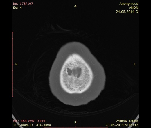
Достоверно признаков поражения костей черепа не нашла.
"Предоставляя весь смысл и совершенство в распоряжение одного только Бога, вы избавляете себя от бездны хлопот." Джон Уитборн.
Если есть возможность при миеломе делайте не КТ - а МРТ с контрастом или ПЭТ-КТ.
Инфильтративного характера мтс в костях по КТ можно не уведеть (если нет явного солидного компонента с деструкцией костных платинок). К тому же оценить динмику на фоне лечения (если нет явных солидных очагов) будет невозможно :(
http://myeloma.org/ROOT/ArticlePage.action?tabId=1&menuId=149&articleId=2944&aTab=-1&gParentType=menuitem&gParentId=149&parentIndexPageId=48&parentCategoryId=36
http://myeloma.org/pdfs/IMWG_consensus_imaging.pdf
http://onlinelibrary.wiley.com/store/10.1111/bjh.12007/asset/bjh12007.pdf?v=1&t=hvlcmg0f&s=005d97cef4e01aed099bed230fe350b2ebc7cfff
http://www.ncbi.nlm.nih.gov/pmc/articles/PMC1665309/
http://www.ncbi.nlm.nih.gov/pmc/articles/PMC2727965/
Небольшая выдержка:
As described above, direct assessment of bone marrow plasma cell infiltra- tion as afforded by MRI is possible before bone lytic lesion (s) appear on WBXR and/or CT (Baur-Melnyk et al, 2005). Therefore, MRI is more sensitive than WBXR in the early detection of MM-related bone destruction. Moreover, MRI is the gold standard technique for the assessment of myeloma- tous involvement of the spine, for several reasons. Firstly, it provides excellent imaging due to its improved sensitivity over conventional radiography (Lecouvet et al, 1999). Sec- ondly, it accurately shows the presence of any spinal cord and/or nerve root compression, and enables the recognition of soft tissue masses (Joffe et al, 1988). Thirdly, MRI can predict the risk of vertebral fracture, even though it does not help in predicting the level of fracture. It has been reported that the risk of vertebral collapse is 6-fold to 10-fold higher in patients with more than 10 FLs on MRI in comparison with those with a normal bone marrow pattern or <10 FLs (Lecouvet et al, 1997, 1998a). Fourthly, it is the best tool for distinguishing between benign and malignant osteoporosis- induced vertebral fractures. This is of great help in clinical practice because the appearance and distribution of vertebral fractures is similar between patients with benign osteoporosis and those with MM. Fifthly, MRI can accurately evaluate the percentage of vertebral height loss before percutaneous vertebroplasty or kyphoplasty (Van Gelderen et al, 1997; Diamond et al, 2004). Finally, MRI allows us to detect complications of the disease, such as soft tissue amyloid deposits, and is the standard technique to be used in the diagnosis of avascular necrosis of the femoral head (Lafforgue et al, 1993).
Выпускник. Таких миелом насчитал более десятка. Ну отметьте пациенту пазухи, похоже там все проблемы.
Всем спасибо.