Radiology Key.
Evaluation and Imaging Features of Benign Breast Masses
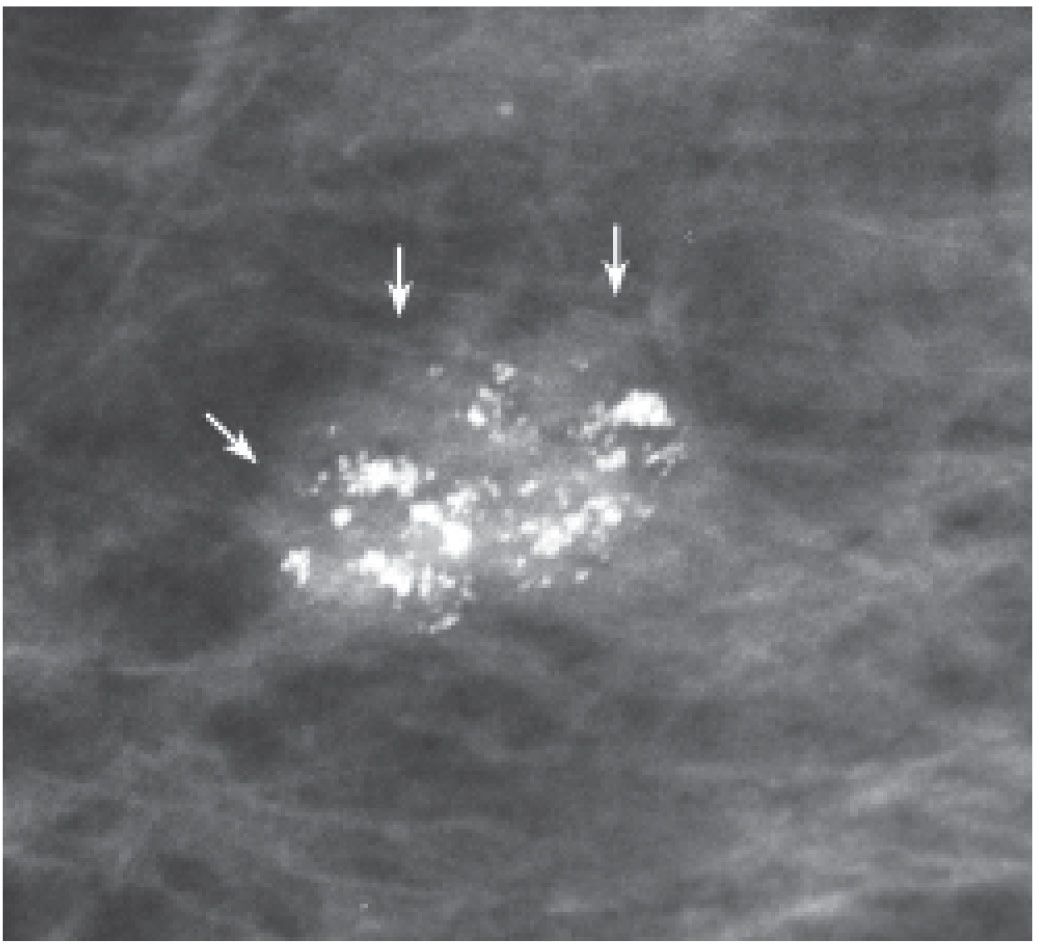
• Фиброаденома
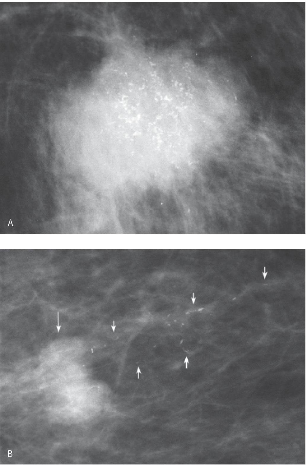
Invasive ductal carcinoma, not otherwise specified (NOS), with DCIS.
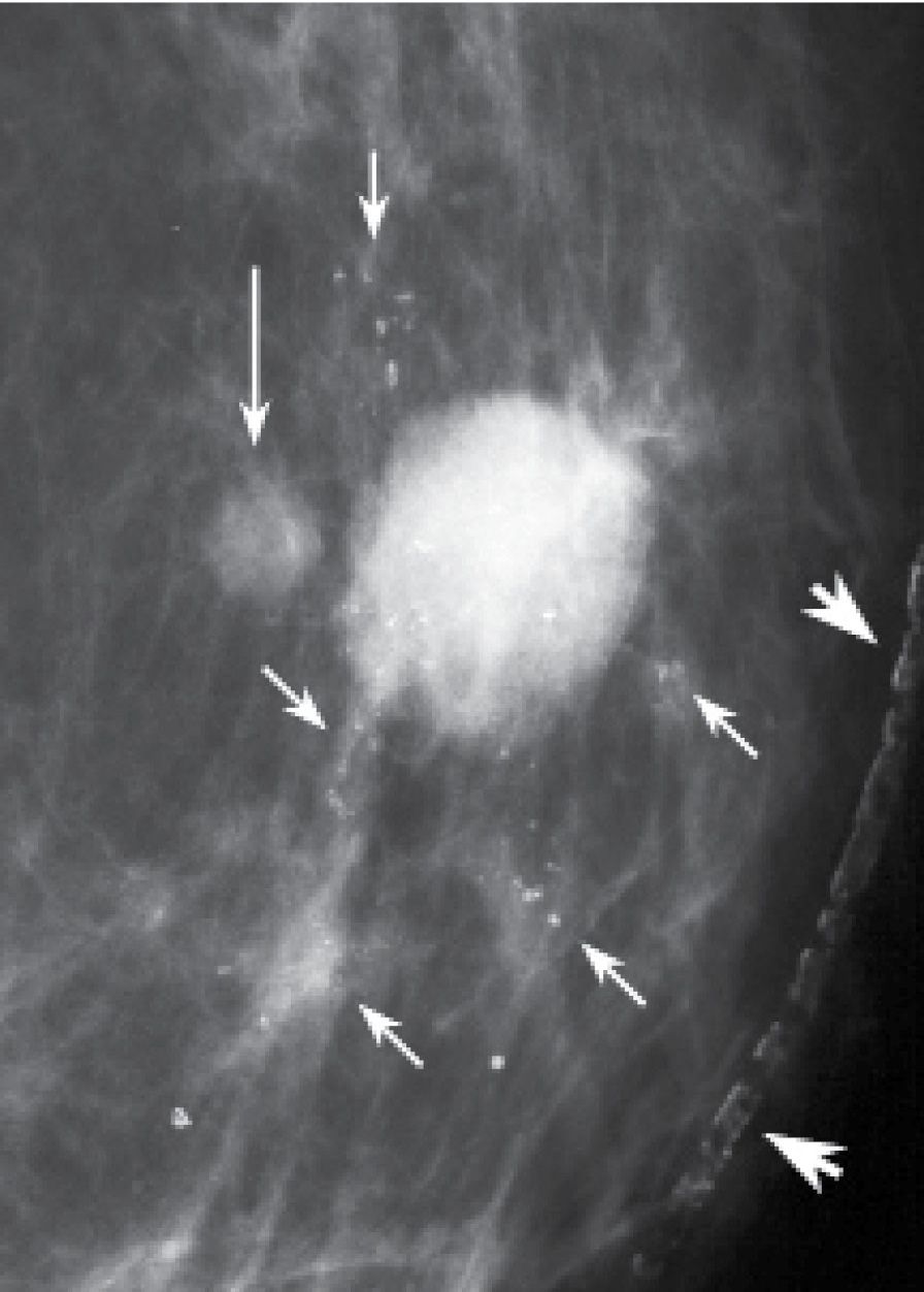
Мультифокальная, инвазивная протоковая карцинома
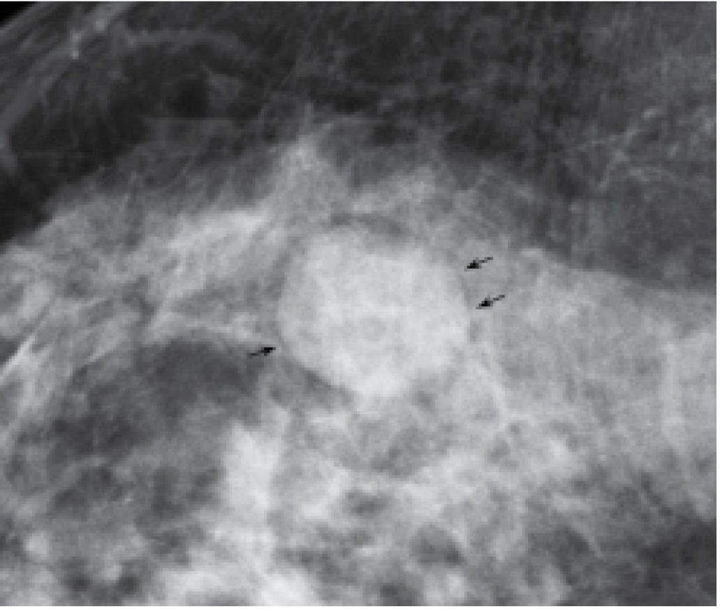
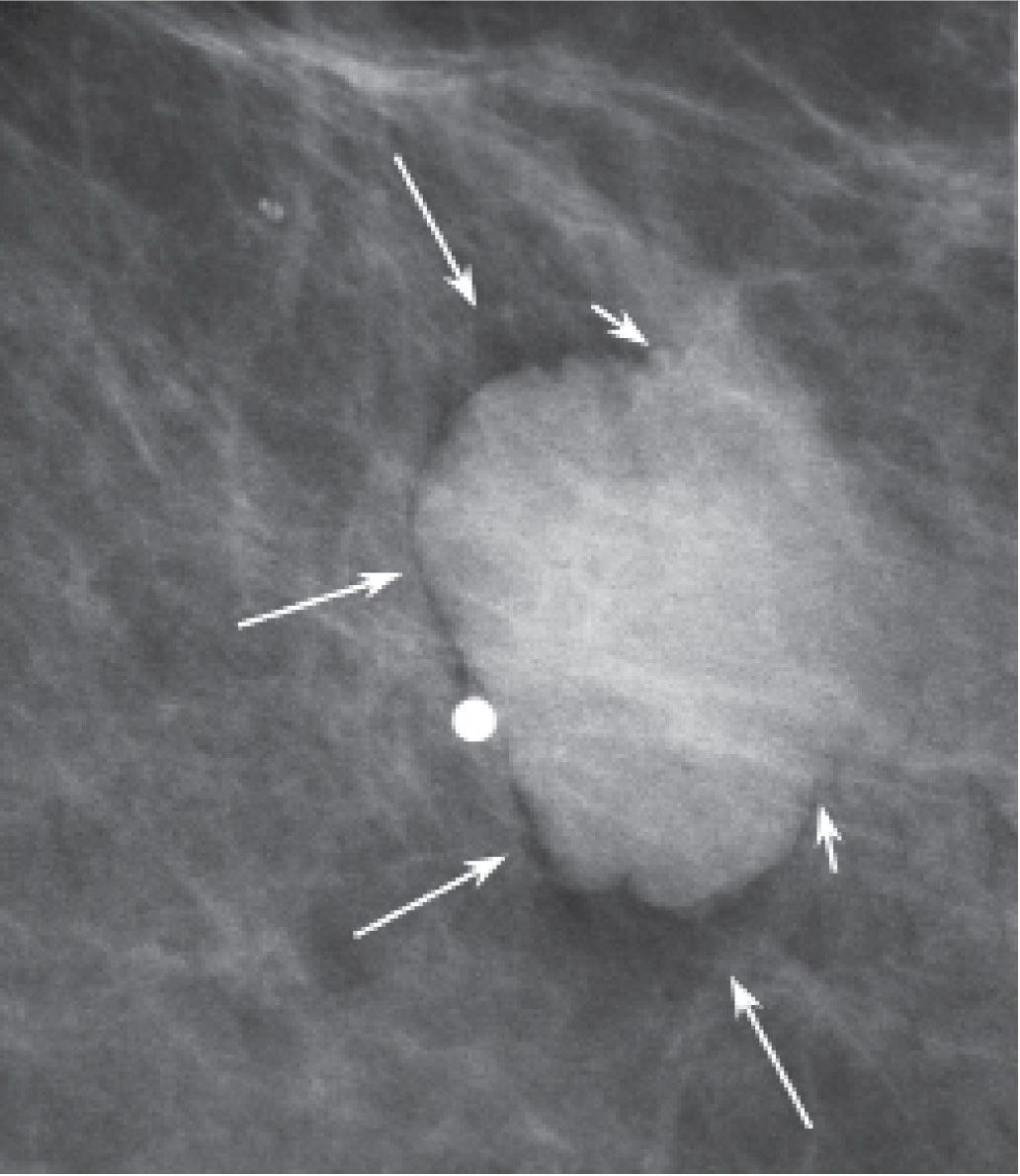
«Halo знак,» киста.
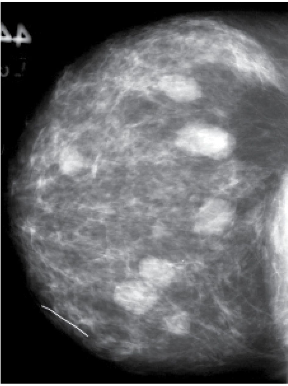
Фиброаденомы.
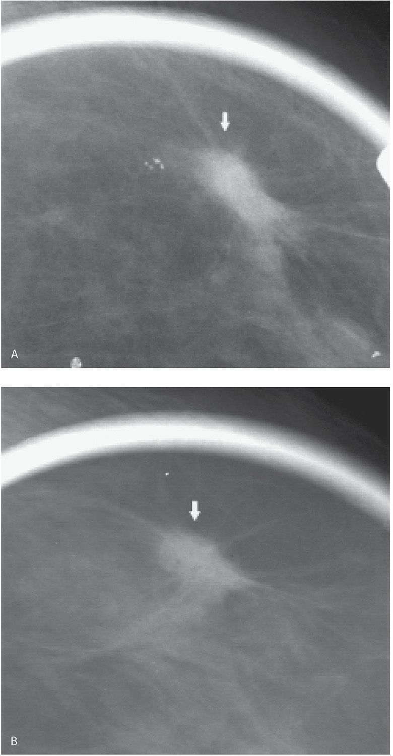
Tubular carcinoma.
Skin lesions.
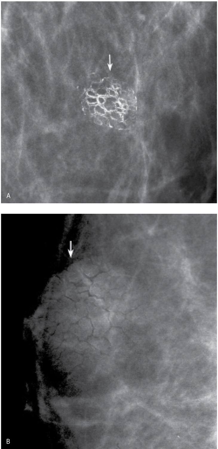
Себорейный кератоз.
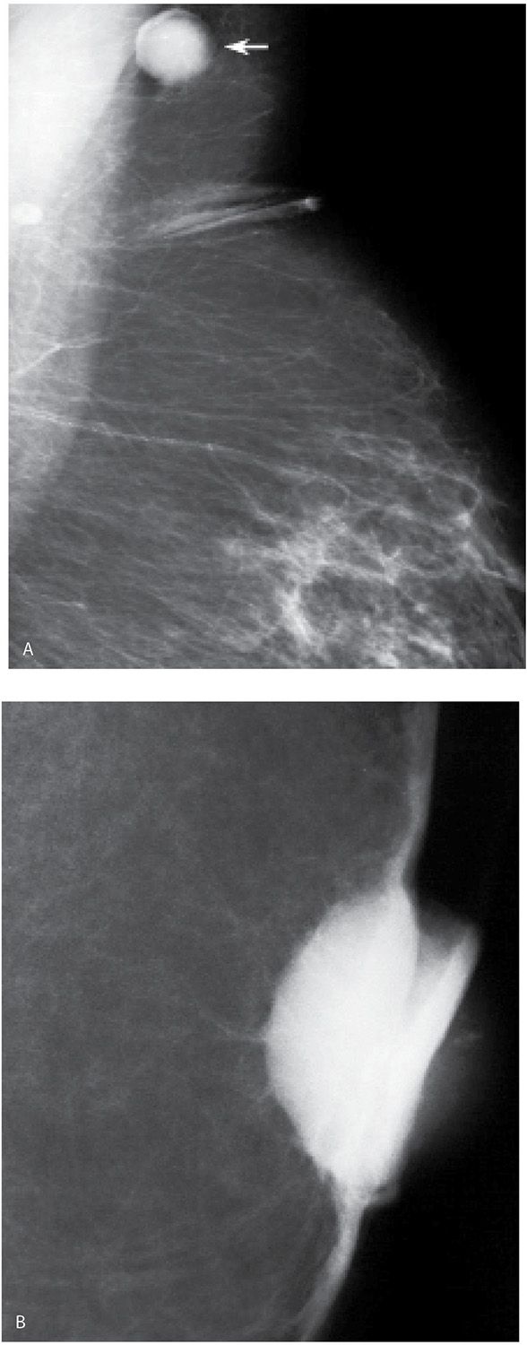
Sebaceous cysts.
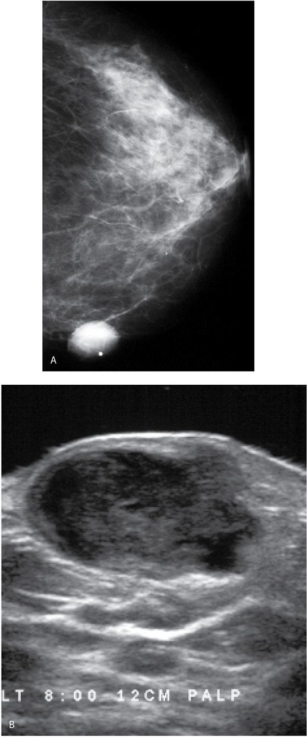
Sebaceous cysts.
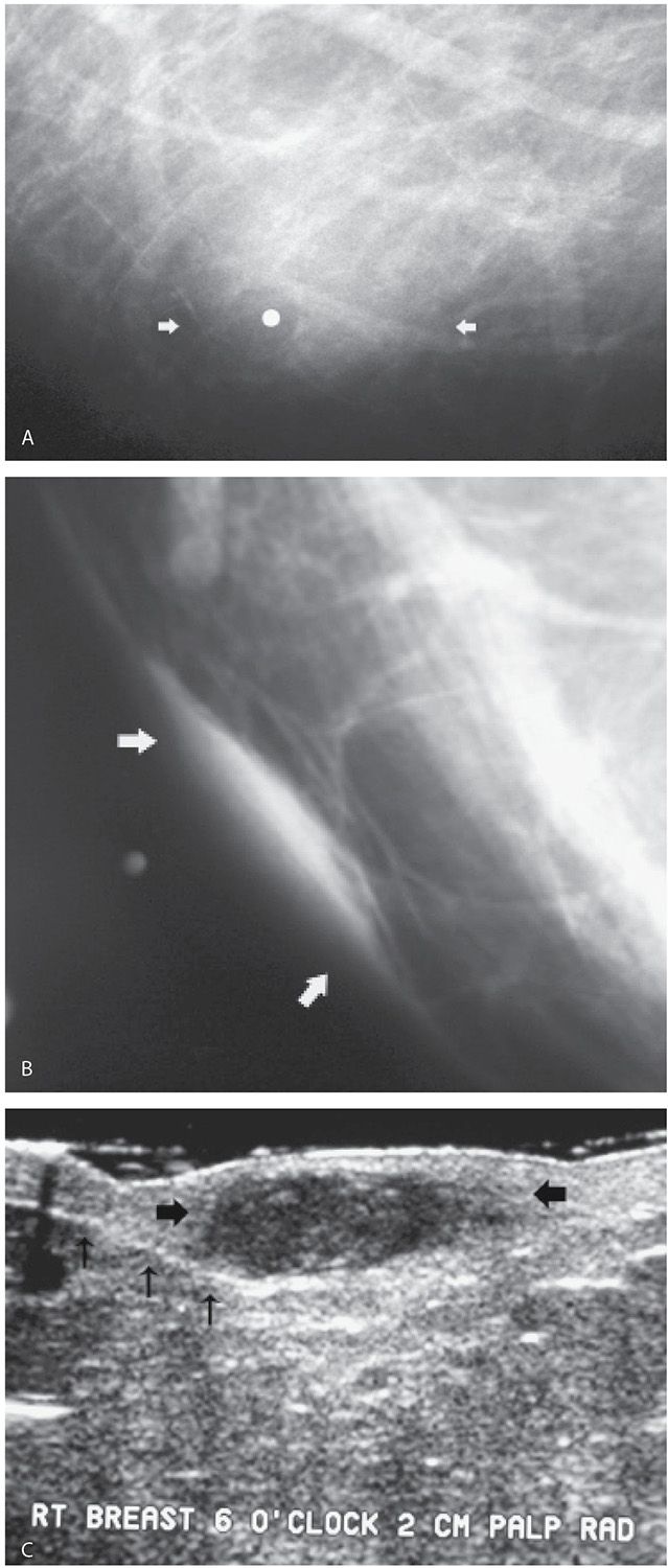
Sebaceous cysts.
https://radiologykey.com/evaluation-and-imaging-features-of-benign-breast-masses/

The Altered Breast
Маркеры биопсии
Изменения, связанные с биопсией, левая грудь.
Biopsy-related fat necrosis.
Evolution of biopsy changes.
Гематома
Гематома
Гематома, эволюция.
https://radiologykey.com/the-altered-breast-2/
Интервенционные процедуры
Nipple discharge.
Establishing accurate cannulation.
Папилломы.
https://radiologykey.com/interventional-procedures-2/
Оценка и визуализация злокачественных образований.
Skin dimpling, invasive ductal carcinoma, NOS, well-differentiated
Nipple inversion and retraction, focal peau d’orange
Skin dimpling and retraction, invasive ductal carcinoma NOS, moderately differentiated.
Изъязвление кожи, инвазивная проксимальная карцинома NOS, умеренно дифференцированная
Necrotic breast, invasive ductal carcinoma NOS, locally advanced with metastatic disease to the contralateral axilla.
https://radiologykey.com/evaluation-and-imaging-features-of-malignant-breast-masses/
Breast Calcifications
Rod-like calcifications.
Resolving arterial calcifications.
https://radiologykey.com/breast-calcifications/
Miscellaneous Mammographic Findings
Эффект радиационной терапии.
Эффект радиационной терапии.
Гранулематозный мастит.
https://radiologykey.com/miscellaneous-mammographic-findings/
Магнитно-резонансная томография груди
Bloom artifact resulting from ferromagnetic objects
Non–fat-suppressed T1-weighted images done using a 2-mm slice thickness.
https://radiologykey.com/breast-magnetic-resonance-imaging/
Мужская грудь
Гинекомастия симметричная.
Гинекомастия асимметричная.
https://radiologykey.com/the-male-breast-3/
Интересные и трудные случаи.
https://radiologykey.com/difficult-cases-6/
Отображение увеличенного пятна влево.