КСС. Череп. Sinus Pericranii. Идиопатический внутричерепной гиперостоз. Cisterna Magna. +

Селшук Пекер, Х. Ибрахим Сан, Сердар Озген, Озлем Курткайя, М. Некметтин Памир

Институт неврологических наук Мармарского университета, Стамбул, Турция

Sinus Pericranii, хирургия, сосудистая патология.

Sinus Pericranii - флюктуирующая сосудистая опухоль с четкими границами, расположенная под кожей черепа. Она сообщается непосредственно с внутричерепной венозной системой. Данный вид патологии наиболее характерен для детской возрастной группы.

По мнению Marras [5], первое упоминание о развитии подобного состояния вследствие перелома костей черепа приходится на 1760 год и, вероятно, связано с именем Percivall Pott. В 1845 Hecker вводит новый термин для обозначения данного феномена: "varix spurius circumscriptus venae diploicae frontalis" [2].

В данном сообщении мы приводим описание Sinus Pericranii у молодого человека.

Описание случая

Молодой мужчина (21 год) поступил с жалобами на головную боль и припухлость мягких тканей в правой лобной области, которые наблюдались в течение года. Kакие-либо сведения о травме головы в анамнезе отсутствовали.

При клиническом осмотре было обнаружено мягкое, податливое и не пульсирующее объемное образование в правой лобной области размером 5´3 см. Состояние кожных покровов над ним было в норме. Размер образования увеличивался при выполнении пациентом движения согласно тесту Valsalva. Неврологическое обследование не выявило каких-либо патологических изменений.

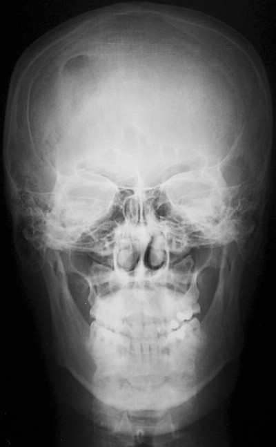
Обзорные рентгеновские снимки черепа свидетельствовали о наличии участка остеолизиса со склеротическими краями в правой лобной области (рис.1).

Рис.1. Обзорная рентгенограмма черепа: область остеолизиса лобной кости справа.

По данным компьютерной томографии (КТ) на поверхности лобной кости отмечалась небольшая эрозия (рис.2).

Рис.2. КТ в режиме костного окна свидетельствует о наличии эрозии на поверхности кости.

Обследование с помощью магнитно-резонансной томографии (МРТ) выявило наличие образования, которое было гипо- и гиперинтенсивным на Т1- и Т2-взвешенных изображениях соответственно. При использовании Gd-DTPA оно характеризовалось неоднородным усилением (рис.3).

Рис.3. Сагиттальное Т1-взвешенное изображение с усилением Gd-DTPA.

Был поставлен следующий клинический диагноз: Sinus Pericranii.


Хирургическое лечение

Под общим наркозом был выполнен дугообразный разрез кожи выше линии волос. Опухоль располагалась под надкостницей.


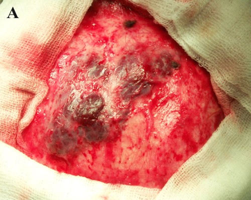
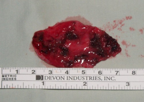
Выявленная патология. По данным микроскопического исследования опухоль состояла из множества венозных сосудов с отсутствующим мышечным слоем, выстланных обычными эндотелиальными клетками. В ряде областей внутри опухоли была обнаружена фиброзная ткань.

Обсуждение

Sinus Pericranii - это скопление венозных сосудов без мышечного слоя или венозная гемангиома, плотно прилегающая к наружной поверхности кости черепа и сообщающаяся внутри черепа непосредственно с синусом твердой мозговой оболочки через диплоические вены [2].

В 1850 г. Stromeyer описал этот вид патологии, как "субпериостальную кисту черепа, заполненную кровью и сообщающуюся с синусом твердой мозговой оболочки", и предложил термин Sinus Pericranii [8]. Медицинская литература содержит множество терминов для обозначения данной нозологической единицы: простое варикозное расширение (varix simplex), гроздевидное варикозное расширение (varix racemosus), варикозный герниоз (varix herniosis), варикозная аневризма (cirsoid aneurysm), венозная ангиома (venous angioma), варикозно расширенная вена (varix cirsoideus), остеососудистый свищ (fistule osteovasculaire), венозная опухоль костей черепа, варикоз вен черепа [7].

Причина появления этого образования пока не ясна. Обычно Sinus Pericranii считается врожденной патологией, хотя есть предположения, что травма также может быть возможным причинным фактором, по крайней мере, в ряде случаев [4]. Общепринятым критерием для установления первопричины данной патологии является тип ткани, выстилающей мешок. Эндотелиальная выстилка характерна для врожденного Sinus Pericranii, тогда как приобретенное образование имеет выстилку из соединительной ткани [7]. Микроскопическое исследование опухоли в нашем случае свидетельствовало о наличии эндотелиальных клеток. Таким образом, мы пришли к выводу о врожденном характере Sinus Pericranii у нашего больного.

Обычно Sinus Pericranii располагается по соседству с синусами твердой мозговой оболочки. Есть несколько сообщений о его латеральной локализации в лобной или лобно-височной областях [7].

Как правило, эта патология не проявляется какими-либо симптомами. Характерной чертой данного образования является увеличение его размера при повышении внутричерепного давления, обусловленного плачем, выполнением движения согласно тесту Valsalva или вынужденным положением [1]. Размер опухоли уменьшается при непосредственном надавливании или подъеме головы. Это патологическое состояние может развиться в любом возрасте. Однако большинство пациентов обычно моложе 30 лет. Соотношение между больными мужского и женского пола составляет 2:1 [1].

Клинический диагноз Sinus Pericranii может быть поставлен на основании появления мягкого флюктуирующего образования, локализующегося возле синуса твердой мозговой оболочки и увеличивающегося в размере при выполнении движения в соответствии с тестом Valsalva [2]. Данные обзорного рентгенографического исследования могут свидетельствовать о наличии гипоплотной области, расположенной ниже опухоли. КТ с использованием костных окон позволит выявить четко выраженное остеолитическое поражение. Что касается нашего больного, то данные КТ исследования указывали на наличие небольшой эрозии на поверхности лобной кости. Кроме того, может быть виден отек мягких тканей. МРТ дает информацию о содержимом синуса на основании характеристик сигнала и взаимосвязи между поражением и подлежащим синусом [4]. Sadler [6] описал больного, обследованного с помощью МРТ, данные которой указывали на поражение, характеризующееся сигналом со смешанной интенсивностью. Это было обусловлено турбулентным кровотоком. Для определения взаимосвязи между образованием и синусами твердой мозговой оболочки можно применять МР-венографию [1].

Несмотря на данные о возможном спонтанном регрессе Sinus Pericranii [2], в целом, он имеет тенденцию к постепенному росту [7]. У большинства больных какие-либо симптомы отсутствуют. Таким образом, основной целью хирургического лечения является устранение косметического дефекта. Кроме того, удаление образования позволит предотвратить сильное кровотечение и возможность травматической воздушной эмболии. Хирургическое лечение заключается в простом удалении опухоли и блокировании или подавлении коммуникантных вен. У нашего больного коммуникантные диплоические вены были заблокированы с помощью костного воска. Ряд авторов сообщает о применении обширной краниотомии или краниоэктомии в целях удаления данной мальформации [5]. В литературе не описаны случаи, когда окклюзия этого патологического пути привела к развитию внутричерепной венозной гипертензии [3]. К возможным осложнениям данного хирургического вмешательства относятся воздушная эмболия и кровотечение.

ЛИТЕРАТУРА

  1. Azusawa H., Ozaki Y., Shindoh N., Sumi Y. Usefulness of MR venography in diagnosis sinus pericranii: Case report. // Radiation Medicine. - 2000. - Vol.18 (4). - P. 249-252.
  2. Bollar A., Allut A.G., Prieto A., Gelabert M., Becerra E. Sinus pericranii: radiological and etiopathological considerations. // J. Neurosurg. - 1992. - Sept. - Vol. 77. - P. 469-72.
  3. Desai K., Bhayani R., Goel A., Muzumdar D. Sinus pericranii in the frontal region: A case report. // Neurology India. - 2001. - Sept. - Vol. 49. - P. 305-7.
  4. Jung S., Lee J.K., Kim S.H., Kim J.H., Kang S.S., Lee J.H. Parietal sinus pericranii. // Surg. Neurol. - 2000. Sept. Vol. 54(3). - P. 270-3.
  5. Marras C., McEvoy A.W., Grieve J.P., Jager H.R., Kitchen N.G., Villani R.M. Giant temporooccipital sinus pericranii: A case report. // J. Neurosurg. Sci. - 2001. - Jun. - Vol. 45(2). P. 103-6.
  6. Sadler L.R., Tarr R.W., Jungreis C.A. Sinus pericranii: CT and MR findings. // J. Comput. Assist. Tomogr. - 1990. Vol. 14. - P. 124-7.
  7. Spektor S., Weinberger G., Constantini S., Gomori J.M., Beni-Adani L. Giant lateral sinus pericranii. // J. Neurosurg. - 1998. - Jan. - Vol. 88. - P.145-7.
  8. Stromeyer L. Ueber Sinus pericranii. .// Dtsch. Klin. - 1850. - Vol. 2. - P. 160-1.
Катенёв Валентин Львович аватар
Не на сайте
Был на сайте: 5 лет 4 месяцев назад
Зарегистрирован: 22.03.2008 - 22:15
Публикации: 54876

Sinus Pericranii

Приложения: 
111.s.slayd114.jpg112.s.slayd115.jpg114.s.slayd117.jpg115.s.slayd118.jpg
Катенёв Валентин Львович аватар
Не на сайте
Был на сайте: 5 лет 4 месяцев назад
Зарегистрирован: 22.03.2008 - 22:15
Публикации: 54876
Катенёв Валентин Львович аватар
Не на сайте
Был на сайте: 5 лет 4 месяцев назад
Зарегистрирован: 22.03.2008 - 22:15
Публикации: 54876
алкс аватар
Не на сайте
Был на сайте: 8 лет 4 месяцев назад
Зарегистрирован: 24.10.2012 - 22:55
Публикации: 2915

"Таким образом, основной целью хирургического лечения является устранение косметического дефекта."

Два случая лично- никто и не пытался оперировать. Достаточно разъяснения патологии больному.

Катенёв Валентин Львович аватар
Не на сайте
Был на сайте: 5 лет 4 месяцев назад
Зарегистрирован: 22.03.2008 - 22:15
Публикации: 54876

Идиопатический внутричерепной гиперостоз

La Hiperostosis Craneal Interna Idiopática es un hallazgo muy frecuente que se puede observar en las imágenes de Tomografía Computarizada y Tomografía por Resonancia Magnética craneoencefálicas, de personas mayores. Desde el punto de vista morfológico se caracteriza por un engrosamiento del díploe  de algunos huesos del cráneo, focal o generalizado, y distribución simétrica con respecto a la línea media. Es una alteración muy conocida que aparece en mujeres mayores de 60 años y  fue descrita por primera vez, por Morgagni en el año 1719 en un grupo de mujeres que también presentaban obesidad e hirsutismo. La etiología es desconocida, aunque predominan los autores que la consideran de origen hormonal.

Nosotros pensamos que el fenómeno de hiperostosis tiene una función protectora del tejido cerebral. Envuelve al parenquima encefálico con suma delicadeza, en aquellas pacientes mayores de 65 años que sufren atrofia cerebral importante. Con eso el encéfalo queda como protegido en un estuche  entre algodones. Nunca se han encontrado lesiones cerebrales  en las necropsias de pacientes con hiperostosis frontal interna.  La disminución de los espacios libres intracraneales evita que los movimientos bruscos, como un simple traspiés, puedan provocar elongación de las venas y desencadenar una hemorragia. A lo largo de los años hemos comprobado que las mujeres mayores, con hipersostosis craneal, no sufren hematomas subdurales, como los varones.  Por el contrario las mujeres mayores que no padecen atrofia cerebral, tampoco llegan a desarrollar Hiperostosis Craneal. Este hallazgo demuestra que la causa que desencadena la hiperostosis sería la atrofia cerebral que pondría en marcha el proceso de osteogénesis intracraneal. Este fenoméno ya está descrito en los niños con hidrocefalia congénita que luego se corrige quirúrgicamente.
.
The Hyperostosis Frontalis Interna is a benign  process which concerns the diploe and the innner face of supratentorial cranial vault especially the frontal bone. Generally is bilateral and does not affect the sagittal sinus sulcus. The first description was performed by Morgagni, in 1719, in female patients who also had obesity and hirsutism. The etiology is controversial, however predominantly authors consider hormonal origin. We think that brain atrophy would trigger the process of osteogenesis, responsible for cranial hyperostosis in women, as a protective mechanism of the brain, that would trigger a hormonal stimulus.


D%C3%ADploe+1.png


FIGURA 1) Los huesos planos del craneo estan constituidos por dos capas de hueso cortical compacto, con unos valores de atenuación de 1000 UH, que se conocen como la Tabla Externa y la Tabla Interna. Entre ambas, se encuentra el Díploe, una tupida malla de tejido esponjoso delimitado por trabéculas y cavidades que contienen la médula roja hematopoyética y tejido adiposo. Con la edad pueden producirse algunas variaciones de esta arquitectura. 
 
(The flat skull bones are composed of inner and outer layers  formed by compact bone. Both are separated by trabeculae and marrow cavities. The marrow is still relatively cellular with little fat accumulation. It has also many blood-forming cell).


1-A) Funciones del hueso Craneal: L os huesos del cráneo tienen varias funciones. De todas ellas, la más importante es proteger al  parénquima encefálico de los traumatismos y desplazamientos bruscos. Es, por tanto, un tejido vivo que está en continuo cambio, para intentar solucionar los problemas que le plantea el envejecimiento de las personas. La segunda función que tiene encomendada es proteger la médula hematopoyética que se acumula entre sus cavidades.
 
(Skull Bone Functions: Protection, support, movement, mineral storage, and blood cell production are all functions of bone)
 
1-B) Producción de Hueso: El fenómeno de producción de hueso nuevo se denomina Osteogénesis, y es un  fenómeno bastante frecuente en los huesos del cráneo, porque se puede comprobar en las exploraciones de TC y TRM craneoencefálicas realizadas a pacientes mayores, especialmente las mujeres. 

Osteogenesis:

Intramembranous bone formation in flat skull bones, is the mechanism of bone formation. Connective tissue are replaced by bone. This process takes place in the flat bones of the skull. In a bone we can found:
 
a) The Outer Table: Is formed by dense bone composed of layers of closely packed osteons. It is thick and its contours are smooth.
 
b) The Diploe: It refers to the spongy bone  tissue of the internal part of flat bones. Spongy bone is also referred to as cancellous bone. The mineralized tissue is seen as  trabeculae. Marrow spaces are also present. The diploe protects the red bone marrow only found in hip bones, ribs, sternum, vertebrae, and ends of long bones, in adults.
 
c) The Inner Table: Is formed by dense bone, composed of layers of closely packed osteons. It is thin and wavy.

Según la superficie craneal que abarca la Hiperostosis se distinguen dos tipos bien diferenciados 

A) HIPEROSTOSIS FRONTAL INTERNA:  La hiperostosis frontal interna  se caracteriza por un engrosamiento del díploe del hueso frontal que hace prominencia en la cavidad craneal.  No se extiende más allá de la sutura coronal (flechas). Es la forma más común y se considera una variante anatómica normal.

Hiper+frontal+2.png

FIGURA 2) Hiperostosis frontal interna típica que afecta exclusivamente al hueso frontal. (Typical  frontal hyperostosis that exclusively involves frontal bone).


Hiper+frontal.png

FIGURA 3)  Ventana de Hueso: Hiperostosis frontal interna típica que afecta exclusivamente al hueso frontal. (Bone Window: Typical  frontal hyperostosis that exclusively involves frontal bone).

RM+Hiper+1.png

FIGURA 4) El engrosamiento mamelonado de los huesos del cráneo en la llamada hiperostosis  craneal interna provoca un crecimiento polilobulado del díploe hacia el interior de la cavidad craneal. Es una verdadera osteogénesis de hueso esponjososo neoformado que aparece delimitado por las ondulaciones de la tabla interna. En esta imagen de SE-T1 se aprecia el hueso frontal hiperintenso, por el tejido adiposo de la médula grasa.
 
(Bone osteogenesis which occurs in the so-called internal cranial hyperostosis, causes bone growth mamelonated spongy diploe into the cranial cavity. The newly formed bone appears cancellar, bounded by the undulations of the internal table. It is a true ossification because in this weighted SE image, we can appreciate the T1 hyperintense bone, due to the deposition of adipose tissue in the bone marrow fat).
 

RM+Hiperos+3.png


FIGURA5)  En esta imagen axial FSE-T2  el hueso neoformado de la hiperostosis frontal interna tiene la misma señal que el hueso esponjoso de los parietales donde no se ha producido hiperostosis.
 
(In this axial T2 weighted FSE image, we can see the newly formed bone of the internal frontal hyperostosis has the same signal as the cancellous bone of the parietal bones where there has been no hyperostosis).

RM+hiper+2.png

 
FIGURA 6). En esta imagen axial FSE-Dp  el hueso de la hiperostosis frontal interna tiene la misma señal que el hueso esponjoso. El engrosamiento del hueso no llega a atravesar la sutura coronal.
 
(In this axial Dp weighted FSE image, we can see the newly formed bone of the internal frontal hyperostosis has the same signal as the cancellous bone. Thickening of bone does not reach across the coronal suture).
 
B) HIPEROSTOSIS ENDOCRANEAL DIFUSA:  En la hiperostosis craneal difusa el proceso se extiende más allá del hueso frontal hasta los parietales (Figura 7). Puede simular un proceso patológico. Es más raro.
 
hiper+11.png
 
FIGURA 7) Radiografía lateral de craneo en la que se observan las irregularidades producidas por la neoformación ósea. Simulan algún proceso patológico.
(On this lateral radiograph of skull, irregularities are observed in the inner table. They simulate a disease process).

Hiper.png
 
FIGURA 8) En esta imagen axial  de TC craneoencefalica, correspondiente a la misma persona, se aprecia el parenquima encefálico protegido por los huesos engrosados del craneo.
(On this axial craneoencephalic CT image, corresponding to the same person, we can see the brain parenchyma protected by the skull bones thickened)

Hiper+1.png

FIGURA 9) Con ventana de hueso se puede apreciar el engrosamiento del díploe y el contorno mamelonado de la tabla interna del craneo, hallazgos característicos de Hiperostosis Craneal Difusa Idiopática.
(With bone window you can see the thickening of the diploe and the irregular contour of the inner table of skull, characteristic findings of Diffuse Idiopathic Cranial Hyperostosis)


Hiper+2.png

 FIGURA 10) En esta imagen la línea naranja delimita todo el hueso neoformado.
(In this picture the orange line delineates all the newly formed bone)

Hiper+3.png

FIGURA 11) Límite posterior de la hiperostosis (flechas) que se extiende hasta los huesos parietales.
(Posterior boundary of the hyperostosis (arrows) extending to the parietal bones)

Hiper+4.png

 
FIGURA 12) Festoneado de la tabla interna frontoparietal. (Typical scalloping of the frontoparietal  inner table).


Hiper+5.png


FIGURA 13) Mamelones óseos de hiperostosis craneal interna en un corte más cefálico.
(In this axial CT image we can observe some typicals bone mammelons of internal cranial hyperostosis).


Hiper+atrofia.png

FIGURA 14) Este paciente varón de 78 años tenía una importante atrofia encefálica difusa de predominio frontal. Esta circunstancia provoca que el encéfalo se mueva dentro de una cavidad craneal que le ha quedado grande, por el simple hecho de caminar. Corre más riesgo que otras personas de sufrir un sangrado subdural por desgarro de las venas de la convexidad cerebral. ¿Se podría disminuir el riesgo de alguna forma?
 
(This 78 year old male patient had a diffuse brain atrophy important-dominated front. This circumstance causes the brain to move within the cranial cavity which has been great, by the simple act of walking. Higher risk than others of suffering subdural bleeding by tearing of the veins of the cerebral convexity. Could reduce the risk in any way)
 

Hiper+atrofia+2.png


FIGURA 15) Imagen manipulada con el PAINT. A la imagen anterior se le ha pintado una banda de hueso que simula una hiperostosis frontal interna. No hay duda que el parenquima encefálico queda más sujeto y la posibilidad de que se rompa una vena por un proceso de aceleración brusca, es más difícil.

(Image manipulated  with Windows PAINT program. In the previous image has been painted a strip of bone that simulates an internal frontal hyperostosis. No doubt the brain parenchyma is more subject and the possibility of breaking a vein by a sudden acceleration process is more difficult.)


1) HIPEROSTOSIS CRANEAL DIFUSA.

94 years old, female. She consulted by headaches.


Hiperosotosis.png
 FIGURA 1)


hiperostosis+2.png


 FIGURA 2)

hiperostosis+3.png

  FIGURA 3)

hiperostosis+4.png
 
 FIGURA 4) 

hiperostosis+5.png


FIGURA 5)

 
hiperostosis+6.png
 FIGURA 6)
Катенёв Валентин Львович аватар
Не на сайте
Был на сайте: 5 лет 4 месяцев назад
Зарегистрирован: 22.03.2008 - 22:15
Публикации: 54876

Cisterna Magna

Con el nombre de Cisterna Magna o Cerebelomedular (figuras 1, 2, 3 y 4), se conoce a una parte del espacio subaracnoideo de la fosa posterior craneal donde drena el líquido cefalorraquídeo desde el IV ventrículo, a traves de los agujeros de Luschka laterales y el central de Magendie. No tiene mucha importancia desde el punto de vista anatómico, pero su morfología y el tamaño son tan variables que inducen a confusión, cuando se aprecia de mayor tamaño que el esperado. Cuando este espacio es mas grande de lo normal se denomina Megacisterna Magna. Es una variante anatómica que no tiene significación patológica. El aumento de volumen de la cisterna es secundario  a un defecto congénito en el desarrollo del cerebelo, especialmente el vermis. Nunca debe confundirse con un quiste aracnoideo que sí podría tener consecuencias patológicas. A continuación presentamos varios casos de cisternas magnas, descubiertas de manera fortuita en exploraciones de TC y TRM realizadas por problemas neurológicos que no tenían relación con el tamaño de la cisterna.
 
(With the name of Cisterna Magna or Cerebellomedullary Cistern (Figures 1,2,3 and 4), is known to a part of subarachnoid space of the posterior cranial fossa, where cerebrospinal fluid drains from the fourth ventricle through the lateral holes of Luschka and the central of Magendie. It is not very important from the point of anatomical view, but their morphology and size variables are so confusing when we appreciate it larger than expected.When this space is bigger than normal is called Mega Cisterna Magna. It is a normal anatomic variant without pathological significance. The increased volume of the cistern is due to a congenital defect in the development of the cerebellum, especially the vermis. It wil never to be confused with an arachnoid cyst. Here are several cases of cisterns magna, discovered fortuitously in CT and MRT exams performed by neurological problems that were not related to the size of the cisterns).
 
Magna+0r.png
FIGURA 1) TRM sagital. La Cisterna Magna está delimitada por la superficie inferior del cerebelo, la cara posterior de la médula cervical y la superficie interna del hueso occipital, recubierto éste por la duramadre y la aracnoides.
(MRI saggital.The Cistern Magna is delimited by the lower surface of the cerebellum, the posterior face of the cervical cord and the inner surface of the occipital bone covered by the arachnoid and dura mater membranes).
Magna+00.png

FIGURA 2) Vista de la Cisterna Magna en proyección coronal.

(View of the Cisterna Magna in coronal projection)

Magna+000.png
FIGURA 3) Vista de la Cisterna Magna en proyección axial.
(View of the Cisterna Magna in axial projection)
Magna+0000.png
FIGURA 4) En la mayoría de las personas la Cisterna Magna es tan pequeña que apenas es visible.
(In many people the Cisterna Magna is so small that it is hardly visible).

CASO 1                                                                               

Magna+1.png
FIGURA 1) El tamaño y la morfología de la Cisterna Magna son congénitos, por eso ya puede verse más grande de lo habitual, en exploraciones de TRM realizadas en niños. Flecha roja.
(The size and morphology of the Cisterna Magna are congenital. So that's the cause of it is found larger than usual  in MRI scans performed in children. Red arrow)
Magna+2.png
FIGURA 2) Vista de la Cisterna Magna normal en proyección axial.
(View of a normal Cisterna Magna in axial projection)

CASO 2                                                                             
 

Magna+32.png
FIGURA 1) Vista de la Cisterna Magna normal en proyección sagital.
(View of a normal Cisterna Magna in saggital projection)
Magna+33.png
FIGURA 2) Vista de la Cisterna Magna normal en proyección axial. La existencia de la hoz del cerebelo permite distinguirla de un quiste aracnoideo.
(View of a normal Cisterna Magna in axial projection.The existence of the falx cerebelli can distinguish it from an arachnoid cyst. )
Magna+31.png
FIGURA 3) Vista de la Cisterna Magna normal en una imagen de TC axial. La existencia de la hoz del cerebelo permite distinguirla de un quiste aracnoideo.
(View of a normal Cisterna Magna in a TC axial projection.The existence of the falx cerebelli can distinguish it from an arachnoid cyst. )

 CASO 3                                                                             

Magna+22.png
FIGURA 1) Vista de la Cisterna Magna más grande de lo habitual. ¿Megacisterna Magna?. Normal.
(Sagittal view of the Cisterna Magna larger than usual. Megacisterna Magna?. Normal )
Magna+23.png
FIGURA 2) Megacisterna Magna, normal.
Megacisterna Magna, normal )

CASO 4                                                                              

Magna+41.png
 FIGURA 1) Vista de la Cisterna Magna más grande de lo habitual. Megacisterna Magna, normal.
 (Sagittal view of the Cisterna Magna larger than usual. Megacisterna Magna, normal )
Magna+42.png
FIGURA 2) Megacisterna Magna en una imagen de TRM axial. La existencia de septos meníngeos, y las extensiones laterales en forma de alas, permite distinguirla de un quiste aracnoideo.
(View of the Megacisterna Magna in this MRI axial  image. The existence of meningeal septa, and the lateral extensions in form of wings,  enables us to distinguish it from an arachnoid cyst)

 CASO 5                                                                             
 

Magna+51.png
FIGURA 1) Vista en proyección sagital de una Megacisterna Magna.También aparece aumentada de tamaño la cisterna ambiens. Atrofia vermiana.
(Sagittal projection of a Megacisterna Magna. Also appears enlarged the Ambient Cistern. Vermian atrophy. )
Magna+52.png
FIGURA 2) Megacisterna Magna en una imagen de TRM axial. La existencia de septos meníngeos, y las extensiones laterales en forma de alas, permite distinguirla de un quiste aracnoideo.
(Megacisterna Magna in this MRI axial  image. The existence of meningeal septa, and the lateral extensions in form of wings,  enables us to distinguish it from an arachnoid cyst)
Magna+53.png

FIGURA 3) Megacisterna Magna en una imagen de TC axial.
(Megacisterna Magna in this CT axial  image.)
CASO 6                                                                             

Magna+61.png
FIGURA 1) Proyección sagital de una Megacisterna Magna. Atrofia vermiana.
(Sagittal projection of a Megacisterna Magna. Vermian atrophy. )
Magna+62.png
FIGURA 2) Megacisterna Magna en una imagen de TRM axial. Atrofia Vermiana
(Megacisterna Magna in this MRI axial  image. Vermian Atrophy)

 CASO 7                                                                             

Magna+71.png
FIGURA 1) Proyección sagital de una Megacisterna Magna. Atrofia vermiana.
(Sagittal projection of a Megacisterna Magna. Vermian atrophy. )
Magna+72.png
FIGURA 2) Megacisterna Magna en una imagen de TRM axial. Se aprecia la Hoz del cerebelo que la parte en dos. Atrofia cerebelosa.
(Megacisterna Magna in this MRI axial  image. Cerebellar atrophy. The falx cerebelli)

CASO 8                                                                             

Magna+101.png

FIGURA 1) Proyección sagital de una Megacisterna Magna. Atrofia vermiana.

(Sagittal projection of a Megacisterna Magna. Vermian atrophy. )

Magna+102.png
FIGURA 2) Megacisterna Magna en una imagen de TRM axial. Atrofia cerebelosa. Hoz del cerebelo.
(Megacisterna Magna in this MRI axial  image. Cerebellar atrophy. Falx cerebelli)

 CASO 9                                                                             

Magna+81.png
FIGURA 1) Proyección sagital de una Megacisterna Magna. Atrofia vermiana.
(Sagittal projection of a Megacisterna Magna. Vermian atrophy. )
Magna+103.png
FIGURA 2) Megacisterna Magna en una imagen de TRM axial. Hoz del cerebelo. Atrofia cerebelosa.
(Megacisterna Magna in this MRI axial  image. Falx cerebelli. Cerebellar atrophy)

CASO 10                                                                            

Magna+91.png
FIGURA 1) Megacisterna Magna en una imagen de TC axial.
(Megacisterna Magna in this CT axial  image. )
Magna+92.png
 FIGURA 2) Megacisterna Magna en una imagen de TC axial. Hoz del cerebelo.
(Megacisterna Magna in this CT axial  image. Falx cerebelli )

 CASO 11                                                                            
 

FIGURA 1) Megacisterna Magna en una imagen de TC axial. Septos meningeos. Atrofia cerebelosa
(Megacisterna Magna in this CT axial  image. Meningeal septa. Cerebellar atrophy.)
Magna+111.png

 FIGURA 2) Megacisterna Magna. Septos meningeos. Atrofia cerebelosa
(Megacisterna Magna. Meningeal septa. Cerebellar atrophy.)

Катенёв Валентин Львович аватар
Не на сайте
Был на сайте: 5 лет 4 месяцев назад
Зарегистрирован: 22.03.2008 - 22:15
Публикации: 54876
 (In the middle of the occipital squama is located the external occipital protuberance. It is defined as a prominence about the centre of the outer surface of the squamous portion of the occipital bone, giving attachment to the ligamentum nuchae. Sometimes a painful prominence is detected in the neck, caused by the deposition of calcium salts that form a long spur like a feline's claw. It is a physiological finding that occurs with age and should not be confused with a bone tumor. To  perform a CT scan to clarify the diagnostic doubt is unreasonable).


                                     Eminencia+2.png
 

FIGURA 1) En esta reconstrucción de la superficie craneal, realizada mediante el algoritmo Volume Rendering se aprecia perfectamente la Protuberancia Occipital Externa. Toshiba Aquilion 64 CT Scanner. HUMS.
(In this reconstruction of the cranial surface, using the Volume Rendering algorithm is well illustrated, the external occipital protuberance).


Protuberancia+4.png
 
FIGURA 2) Vista posterior de la Protuberancia Occipital Externa.  
(Posteroanterior perspective of the external occipital protuberance. Toshiba Aquilion 64 CT Scanner).


Protuberancia+5.png
FIGURA 3) Sección sagital, TC Volume Rendering, del cráneo en la que se aprecian perfectamente la Protuberancia Occipital Interna  y la Externa.
(Sagittal section, CT Volume Rendering of the skull, in which are clearly visible  the Internal Occipital Protuberance and the External).
 
Protuberancia+7.png


FIGURA 4) En una radiografía lateral del cráneo tambien se aprecian con nitidez ambas protuberancias.
(In a lateral radiograph of the skull we can be see, clearly, both Protuberances).


Protuberancia+6.png


FIGURA 5) En una exploración de TC craneoencefálica, la protuberancia occipital interna aparece siempre más prominente que la externa.
(In a CT craneoencephalic exam, the internal occipital protuberance is always more prominent than the external).


Protuberancia+3.png


FIGURA  6) En la radiografía lateral del cráneo se aprecian con nitidez el espolón calcareo que se ha formado a partir de la protuberancia occipital .
(In the lateral radiograph of the skull we can be see, clearly, a calcareous spur that has been formed from the occipital protuberance).


Espol%C3%B3n.png


FIGURA 7) El espolón calcáreo se produce por depósito de sales de calcio en la inserción de los músculos del cuello y de la espalda. Es un fenómeno frecuente en otras partes del cuerpo, como el calcáneo o las vértebras (osteofitos).
(The spur is produced by calcareous deposit of calcium salts in the insertion of the muscles of the neck and back. It is a common phenomenon in other parts of the body such as the calcaneus or vertebrae (osteophytes)


Protuberancia++2.png


FIGURA 8) Cuando se forma un espolón, la protuberancia occipital externa aparece siempre más prominente que la externa.
(In a CT craneoencephalic exam, when a bone spur is formed the external occipital protuberance is always more prominent than the internal).


Protuberancia+1.png


FIGURA 9) Aspecto del espolón en un corte más caudal.
(Appearance of the spur in a CT slice more caudal).


Eminencia+1.png


FIGURA 10) Y en otro corte, todavía más caudal.
 (And another slice, still more caudal)


                        MÁS IMÁGENES........................MORE IMAGES

 CASO 1)                                                                                         


Eminencia.png

                                                                 FIGURA 1)
 

Eminencia+2.png

                                                               FIGURA 2)


CASO 2                                                                                   


occipital+1.png
FIGURA 1) En algunas personas muy mayores, 89 años, el espolón puede adoptar una morfologia redondeada.
(In some very old people, 89 years old, the bony prominence may adopt a rounded morphology).


Occipital+2.png


 FIGURA 2) El espolón contiene hueso compacto cortical y hueso esponjoso.
 (The spur contains compact bone cortical and cancellous bone).

Hospital Universitario Miguel Servet. (HUMS) Zaragoza.Spaiñ