Иллюстрации. Инородные тела. Материал университета Аризоны
Figure 28
Figure 29
Figure 30
Figure 31
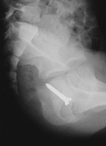
Young child who had placed a screw in her vagina
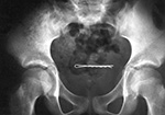
A 2 year-old girl had placed a bobby pin in her bladder (Courtesy George Barnes, MD)
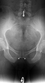
Young man who had inserted a pen refill in his uretra with partial rupture of the urethra and soft tissue gas in the penis.
15 year-old boy with a broken rectal thermometer lying free in the peritoneum. Its orign was unknown. He denied inserting any foreign objects into his rectum or urethra. He had had no treatments or hospitalization since he was one year old. (Courtesty George Barnes, MD)
Figure 32
Figure 33
Figure 34
Figure 35
19 year-old woman with a bobby pin lodged in her uterus. She had attempted to induce an abortion with the bobby pin. A subsequent self-induced abortion was successful. (Courtesy George Barnes, MD)
Young woman with multiple "piercings" including an umbilica ring and labial rings.
18 year-old man with bladder calculi that formed around a wire in his bladder. Six months prior he lost a fine telephone wire in his bladder when he achieved an erection while inserting the wire into his urethra during masturbation. (Courtesy George Barnes, MD)
60 year-old man with a condensed milk can that he had inserted into his rectum. (Courtesy George Barnes, MD)


Figure 36
Figure 37
Figure 38a
Figure 38b
Policeman who somehow lost his traffic wand in his rectum.
Young man with multiple pieces of shrapnel in the hand after gunshot injury.
Young man shot in the knee with multiple bullet debris in the soft tissues about the knee as well as in the knee joint.
Figure 38c
Figure 38d
Figure 38e
Figure 39
Young man who accidently shot himself in the chest and right arm with a shogun. Figure 38c is the scout view from a chest CT. It shows multiple pellets in the right shoulder and right chest wall with contusion of the right upper lobe. A right chest tube, an endotracheal tube, and a nasograstric tube are in place. Figure 38d is a CT image of the lower portion of the chest. There is an inferior pellet in the right ventricle. It had penetrated the right subclavian vein and embolized to the right ventricle. It was visible bouncing in the right ventricle when fluoroscopy was performed during right upper extremity angiography. The pellet was successfully removed at cardiac catheterization. (Courtesy Michael Rosellini, MD)
Transverse ultrasound image of an elderly man with a bullet (arrow) in his liver. He had a history of a gunshot wound many years previously, and the bullet was an incidental finding.
Young man wearing sandals stepped on a nail. The nail penetrated his plantar soft tissues but this radiograph showed it had not entered bone.
Figure 40a
Figure 40b
Figure 41
Figure 42
Underpants in a young woman with metallic material that simulates a pelvic foreign or pelvic surgery.
22 year-old man with large piece of wood (arrows) in his calf after being assaulted with a wooden stake.
17 year-old boy with barnacle fragments in his heel. He had been water skiing when he came into the dock and unexpectedly encountered barnacles, pieces of which lodged into his heel. (Courtesy George Barnes, MD)
Figure 43a
Figure 43b
Figure 44a
Figure 44b
Young man with pieces of glass in his foot after an automobile accident.
Young woman with pieces of glass in her leg after an automobile accident.
Physician who had frequent steroid injections into his left knee prepatellar bursa. He developed a small tender palpable abnormality. At ultrasound two small echogenic lesions (1 and 2) were found. At surgery they were calcific deposits which were removed.
Ultrasound image of a young man who stepped on a broken mesquite tree branch. A large wooden fragment that pierced his foot was removed, but he continued to have pain and swelling over the dorsum of his foot. An echogenic focus was evident on ultrasound (cursors) which proved to be a mesquite thorn at surgery.
Figure 45
Figure 46a
Figure 46b
Figure 47a
Acupuncture needles in the paraspinal subcutaneous soft tissues of 37 year-old Korean woman. The needle fragments were purposefully left in place. (Courtesy Dr. Joseph A. Alvarado)
Elderly man with right hip pain and a draining sinus tract near the right hip. He had a right hip unipolar prosthesis placed several weeks prior in Mexico. Radiographs of the pelvis and right hip (figure 46a) show a unipolar prosthesis as well as a retained surgical sponge (arrow) in the medial aspect of the right acetabulum. The prostheis was removed and replaced with a temporary antibiotic impregnated right hip "prosthesis" (figure 46b) made from antibiotic laden cement and held in place by press fitting and cerclage wires.
Surgical sponges used at University Medical Center, Tucson, AZ. See reference 90.
Figure 47b
Figure 47c
Figure 47d
Figure 48
Surgical needles used at University Medical Center, Tucson, AZ. See reference 90.
Surgical blades used at University Medical Center, Tucson, AZ. See reference 90.
Surgical clips and clamps used at University Medical Center Tucson, AZ. See reference 90.
25 year-old woman who had undergone a Cesarean section in Mexico. She presented with abdominal pain and fever. The abdominal radiograph shows a large complex, partially lucent left flank mass (arrow) with an associated linear density. At surgery there was a retained surgical sponge with surrounding area of granuloma formation, a gossypiboma.
Figure 49
Figure 50a
Figure 50b
Figure 51a
Elderly man with a retained surgical sponge (arrow).
Vertebroplasty cement has embolized to the patient's lungs. This was an incidental findings on a routine chest radiographic series.
21 year-old man who injected himself with metallic mercury.
Figure 51b
Figure 51c
Figure 52
Figure 53
21 year-old man who injected himself with metallic mercury. There are mercury emboli to the lungs and metallic mercury is evident in the antecubital foss of the left elbow. (Figures 51a-c courtesy of Charles A. Rohrmann, Jr, MD. They were originally printed in reference 82).
Trick or treat candy. There are no hidden needles or razor blades. The candy and the apple (round density) where eaten with much enjoyment.
Axial three-dimensional MR arthrogram of the left shoulder in a 40 year-old man as follow-up from prior rotator cuff repair. The arrows point to areas of signal void from presumed tiny metallic deposits in the shoulder from the surgery. Shoulder radiographs were normal.
Figure 1
Figure 2
Figure 3
Figure 4
28 year old woman who periodcially swallowed pins and razor blades. The open safety pin in her descending colon passed without mishap.
Small child who swallowed a metal nut
Boxctter in descending colon
15 year-old mentally incapcitated child who periodically swallowed objecs. This radiograph shows a safety pin and key in the jejunum and rubber flexible Khrushchev doll head in the descending colon. It passed without difficulty. (Courtesy of George Barnes, MD)
Figure 5
Figure 6
Figure 7a
Figure 7b
The arrows show a pencil lodged in the ascending colon. It passed without difficulty which is unusual for such an elongated object.
Elderly patient with a coffee cup fragment lodged in the distal esophagus
35 month-old infant who swallowed a quarter
Figure 8a
Figure 8b
Figure8c
Figure 9
Radiographs and images from a barium swallow of a 62 year-old man who accidentally ingested a pill bottle cap while taking his medications. The cap was lodged in his proximal esophagus. It was removed at endoscopy. Figures 8b and 8c were originally published in reference 12.
Metallic mercury in the appendix of an asymptomatic patient from rupture of mercury bag on a Miller-Abbott tube.
Figure 10
Figures 11a and 11b
Figure 11c
Figure 12
72 year-old man with no symptoms. BBs are in the appendix. He used to eat BBs as a child.
Sequential radiographs of a 4 year-old girl who ingested a watch battery show it first in the stomach and later in the transverse colon. The battery passed without complications.
A three year-old boy swallowed four cylindrical magnets which were evident in his stomach. The next day they were in the sigmoid colon and passed with no patient harm.
Soft tissue neck radiograph of a 1 year-old girl who was coughing afer swallowing a red coin-like plastic wafer. The plastic is lodged in her hypopharnyx (arrowheads). She coughed it up a few minutes later. Most plastic objects are not visible on radiographs.
Figure 13
Figure 14
Figure 15
81 year-old man who swallowed a turkey bone (arrows). It is located just posterior to the cricoid cartilage. The bone could not be seen at direct laryngoscopy but was removed at endoscopy.
Xeroradiograph of an elderly man with painful swallowing after eating a bowl of "oxtail soup." There is an impacted piece of bone lying anterior to the 7th cervical vertebra. Note the prominently calcified posterior margin of the circoid bone (arrow). This normal variant should not be mistaken for a foreign body. The impacted bone fragment was removed from the proximal esophagus at endoscopy.
68 year-old man with difficulty swallowing after eating fish. A bone (arrow) had perforated the hypopharyngeal wall and was lodged in the soft tissues of the neck. Indirect laryngoscopy was negative, and the bone was removed at surgery after an unsuccessful endoscopy.
Figure 16a
Figure 16b
Figure 17
36 year-old woman with painful swallowing after eating chicken. A small calcific density (arrowhead) represents a lodged chicken bone. A radiograph after the woman swallowed a barium capsule shows it temporarily lodged at the point of obstruction. The bone was impacted in her proximal esophagus and removed at rigid endoscopy. Figure 16b was originally published in reference 34.
Radiograph of an adult who was in the habit of swallowing rubber gloves. The mottled lucent area (arrow) in the right lower quadrant represents a rolled up rubber glove in the terminal ileum. It has a similar appearance to swallowed drug packets (Figure 24).
Figure 18
Figure 19a
Figure 19b
Figure 20
8 month-old boy with vague respiratory symptoms. Lateral view of the chest shows a thin, twisted metallic foreign body. A piece of wire was removed from his hypopharynx at endoscopy.
2 year-old girl with severe respiratory symptoms. She had a left sided pneumonia and empyema requiring a tracheostomy. For several days the ring-like metallic density noted on her portable chest radiograph was assumed to be associated with her traheostomy. The above lateral and frontal chest radiographs reveal the metallic density is not associated with the tracheostomy. A bing chip was removed at endoscopy.
37 year-old mentally disabled woman who was admitted comatose with a 24-hour history of difficulty breathing. An open safety pin had perforated the wall of her esophagus and penetrated her larynx. It was extracted at laryngoscopy, and she recovered withoout sequelae.
Figure 21a
Figure 21b
Figure 22a
Figure 22b
13 year-old boy with a pin in his trachea. He had been playing with a blowgun and inhaled a corsage pin. It was removed at bronchoscopy (Courtesy of George Barnes, MD)
Whenever there is a plausible history of a foreign body ingestion, the patient should be examined from the base of the skull to the anus. The radiograph on the left (a) shows no opaque body in the chest or stomach of an 8 year-old boy who was reported to have swallowed a foreign object. The radiologist asked for a repeat radiograph (b) to include all the abdomen and pelvis. There is a metallic tweezer in the child's distal small bowel.
Figure 23
Figure 24a
Figure 24b
Figure 24c
Young child with a history of eating sand. (Courtesy George Barnes, MD)
Radiographs of adults who were smuggling drugs. A packet is visible in the transverse colon (arrow in a). Relatively dense packets are visible in the transverse and descending colons in b and c. (Courtesy Charles A. Rohrmann, Jr., M.D., University of Washington, Seattle)
Figure 25
Figure 26
Figure 27
Figure 28
Radiograph of a typesetter shows opaque lead in his distal colon. He had inadvertently ingested high amounts of lead over a period of years while eating his lunch at the plant. He had very high lead levels in his blood, neurologic symptoms, anorexia, and constipation.
3 year-old boy who ingested multiple iron tablets probably containg ferrous gluconate and sulphate salts. He recovered without sequelae. (Courtesy George Barnes, MD)
Radiograph showing bismuth subsalicylate (Pepto-Bismol) tablets in the right lower quadrant producing a pseudo-appendicolith appearance.(Courtesy Charles A. Rohrmann, Jr., M.D., University of Washington, Seattle)
3 year-old girl who ate half melted crayons which formed a bezoar in the stomach. It required surgical removal.
Figures 1a and 1b: tooth anatomy
Figure 2: Stages of dentition
Figure 3: Tooth nomenclature
E - enamel; D - dentine; P - root pulp; R - tooth root; arrows - lamina dura
Mixed dentition in a child with permanent teeth plus retained primary teeth. The + marks developmental follicles for the third molar and the * marks permanent second molars.
WT-wisdom tooth (3rd molar); 2M-second molar; 1M-first molar; 2B- second bicuspid (premolar); 1B-first bicuspid (premolar); K9-canine; LI-lateral incisor; CI-central incisor. The * marks the mandibular canal.
Dental and Mandibular Devices
Figure 4: teeth numbering (Universal/National System)
Figure 5: miscellaneous dental devices
Figure 6: root canal fillings and porcelain denture teeth
Figure 7: cubic zirconia crown and acrylics
Panoramic view of the mandible shows dental plates with screws (A) and bone ligature wires (B). There are multiple dental alloy restorations (amalgam fillings)
There are root canal fillings with denture retention posts (A) and porcelain denture teeth with pins (B).
A coned mandibular view shows a cubic zirconia crown (*). The zirconia is surrounded by porcelain (horizontal arowwheads). In the next tooth there is an endodontic root canal with gutta percha (arrows), an opaque rubber. The light shade of the teeth (vertical arrowheads) shows composite acrylic restoration.
Figure 8: dental overview
Figure 9: Removable partial denture framework (A); dental crowns (caps) (B)
Figure 10: Temporomandibular prosthetic condyle implant (A); orthodontic arch bars (B); porcelain veneer dental crowns (caps) (C); fixation screws (bone screws) (D)
Figure 11: Osseointegrated dental implants with fixed dental bridgework (A); maxillary denture with acrylic teeth
A. Alloy dental restoraration; B. root canal filling with porcelain veneer crowns and posts; C. fixed bridge pontic with abutment crowns; D. dental composite (acrylic) restoration (tooth-colored fillings).
Figure 12: Subperiosteal dental implant with fixed bridge (A); bone plate with screws (B); fixed dental bridgework with root canal fillings (C); fixation wire (D)
Figure 13: Dental implant crown
Figure 14: Zimmer Trabecular Metal Implant
Figure 15: Zimmer Trabecular Metal Implant (on left) and conventional metal implant (on right)
Figure 16: Permanent mandibular retainer
Panoramic view of 38 year-old woman with a permanent (metallic) mandibular retainer.