Менингиома.
С учетом топографии
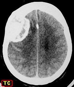
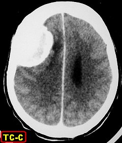
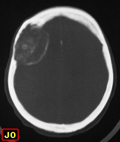
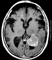
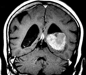
 Meningiomas of cerebral convexityOf falx or parafalcineOf olfactory groove or roof of orbitOf sellar region
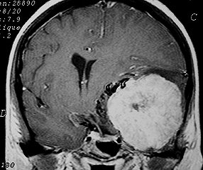
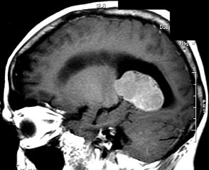
 Of middle fossaOf sphenoid ridgeOf optic nerveOf clivus
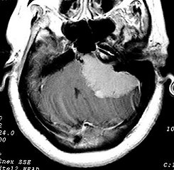
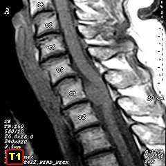
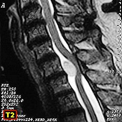
 Of cerebellopontine angleOf tentorium /posterior fossaIntraventricularOf spinal canal
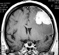
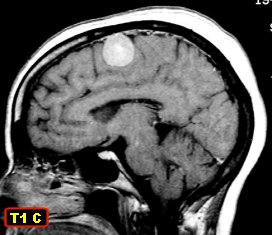
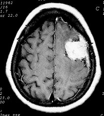
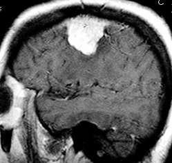
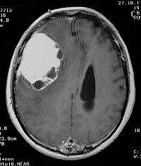
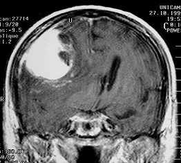
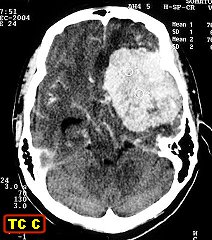
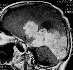
CEREBRAL CONVEXITY
..
....
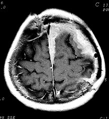
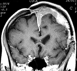
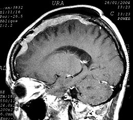
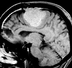
FALCINE / PARASAGITTAL
..
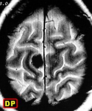
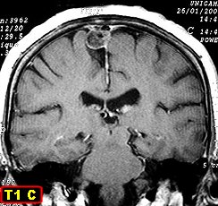
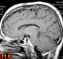
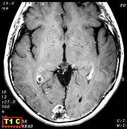
....
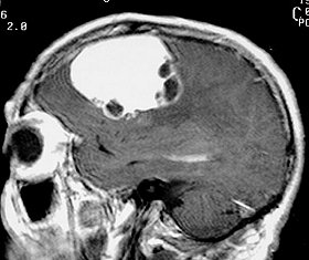
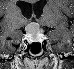
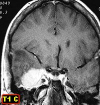
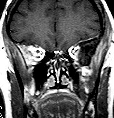
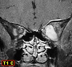
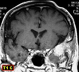
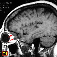
OLFACTORY GROOVE / ORBITAL ROOF
..
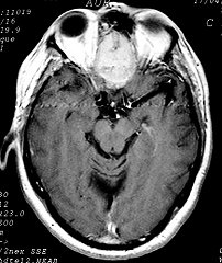
...
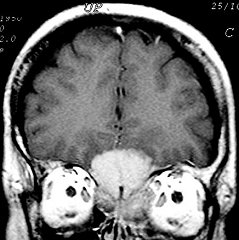
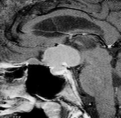
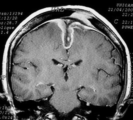
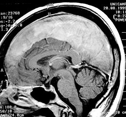
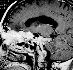
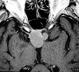
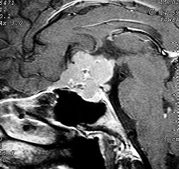
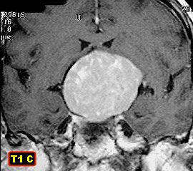
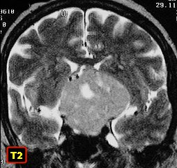
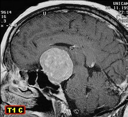
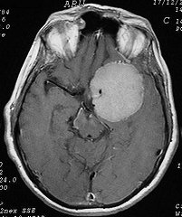
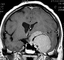
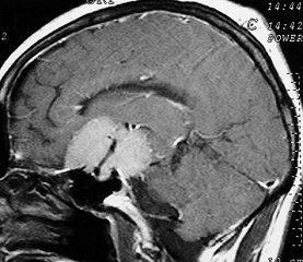
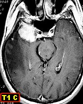
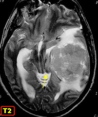
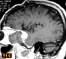
SELLAR REGION
..
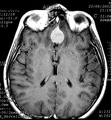
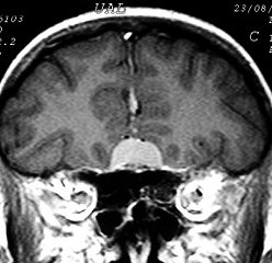
.....
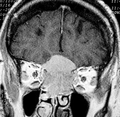
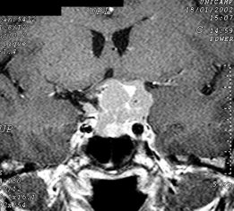
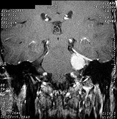
MIDDLE FOSSA
..
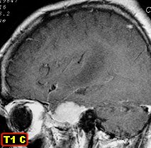
....
SPHENOID RIDGE
..
....
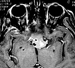
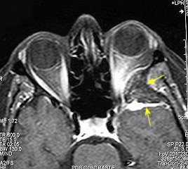
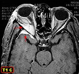
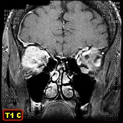
OPTIC NERVE
..
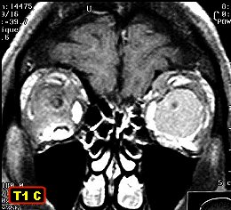
....
CLIVUS
..
....
PETROCLIVAL /
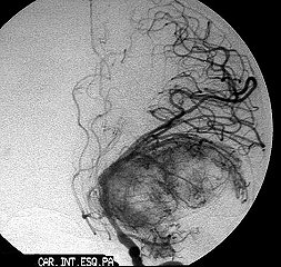
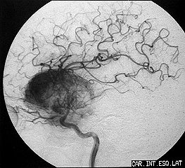
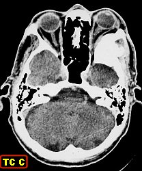
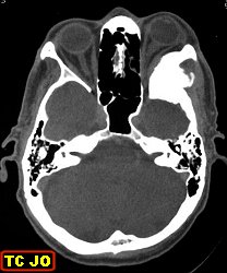
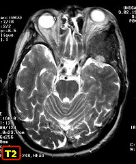
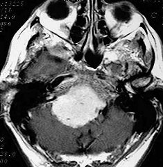
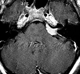
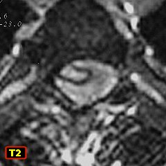
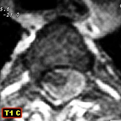
CEREBELLOPONTINE ANGLE
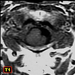
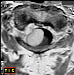
CEREBELLOPONTINE ANGLE
..
....
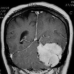
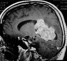
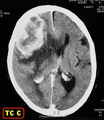
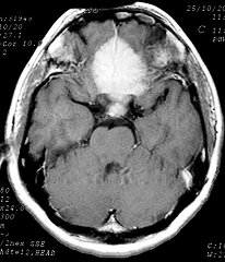
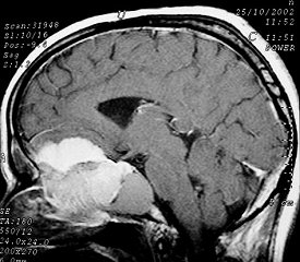
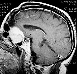
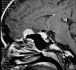
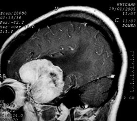
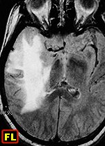
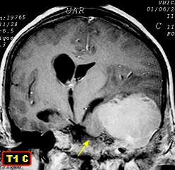
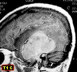
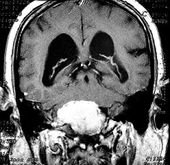
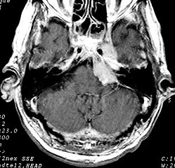
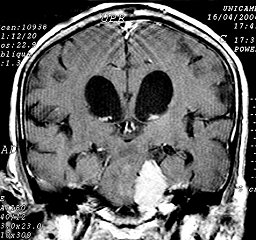
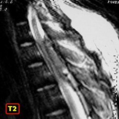
TENTORIUM CEREBELLI / POSTERIOR FOSSA
..
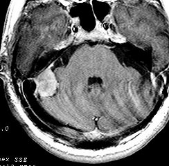
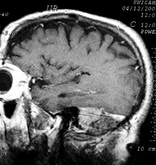
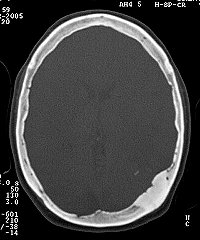
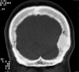
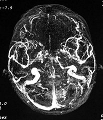
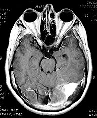
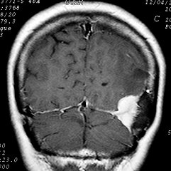
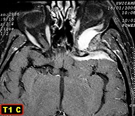
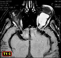
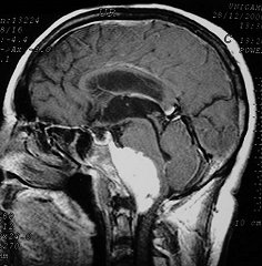
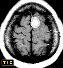
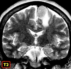
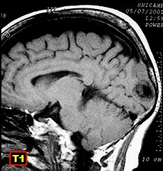
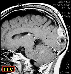
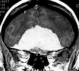
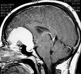
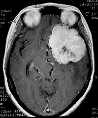
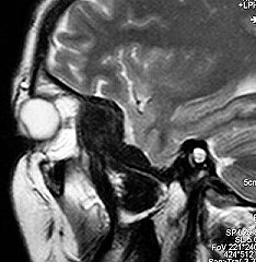
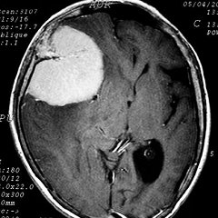
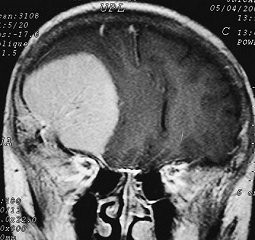
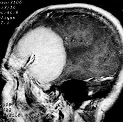
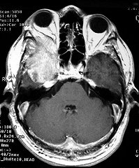
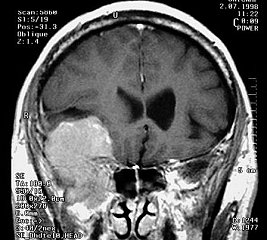
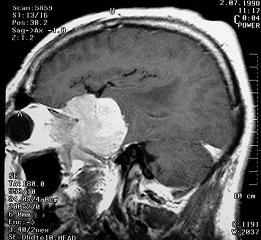
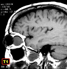
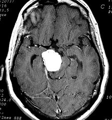
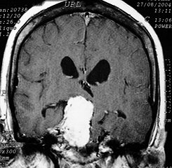
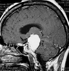
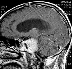
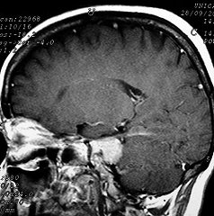
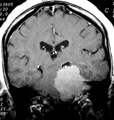
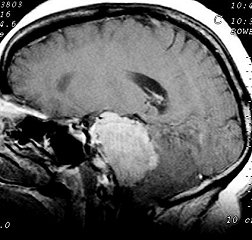
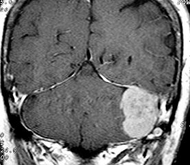
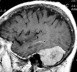
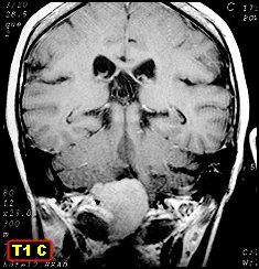
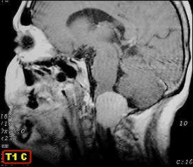
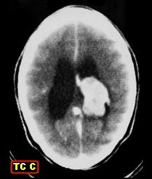
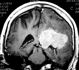
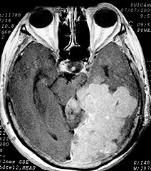
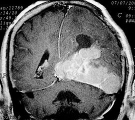
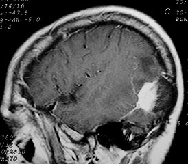 F 48 yr. Meningioma of tentorium infiltrating transverse sinus and occipital bone, with hyperostosis
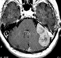
....
INTRAVENTRICULAR
..


....
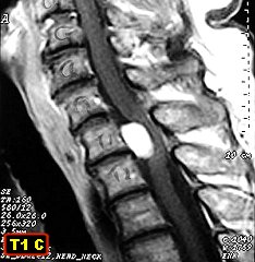
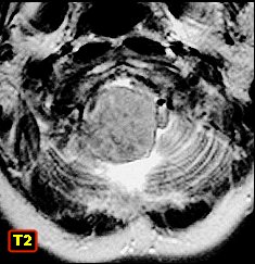
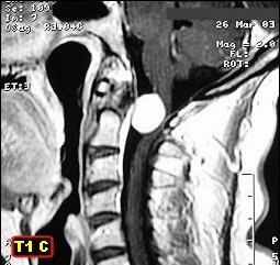
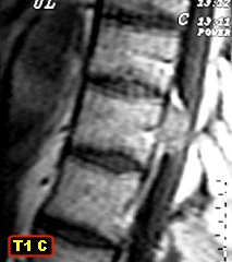
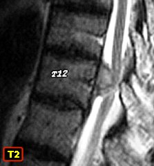
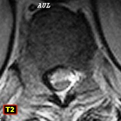
SPINAL CANAL
..
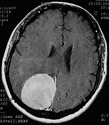
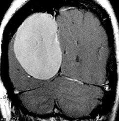
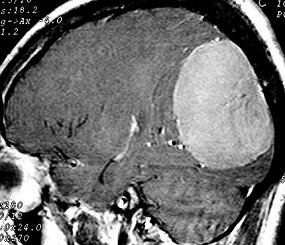
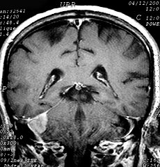
MORE MENINGIOMAS
Fibroblastic
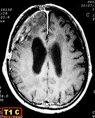
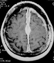
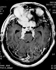
meningioma of left lateral ventricle
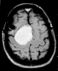
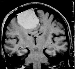
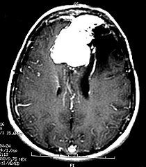
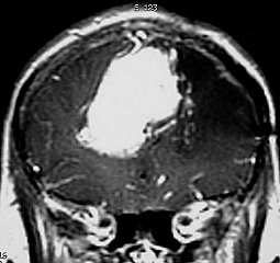
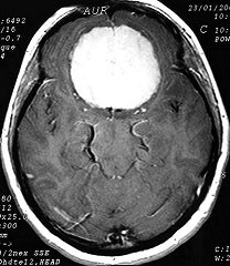
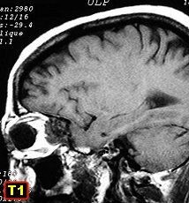
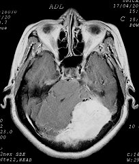
fronto-parietal
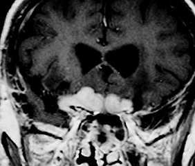
meningioma with hyperostosis and brain compression (MRI Feb 2008)
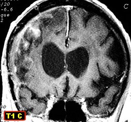
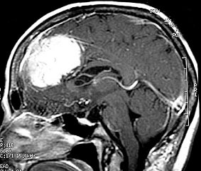
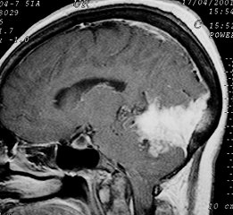
Tumor growth through bone gap and under scalp (MRI Oct 2011)
Гемангиоперицитома
SOLITARY FIBROUS TUMOR OF THE MENINGES
Шваннома
Краниофарингиома
Эпидермоидная / дермоидная кисты
Dermoid cyst (mature cystic teratoma) of sellar region. Spread of lipid droplets through subarachnoid space and lateral ventricle. Text
Опухоли костей
Дифференциальная диагностика
http://www.wjgnet.com/1949-8470/full/v4/i3/75.htm#__sec10