Radiology Key. Врожденные аномалии матки.
https://radiologykey.com/congenital-uterine-anomalies-2/
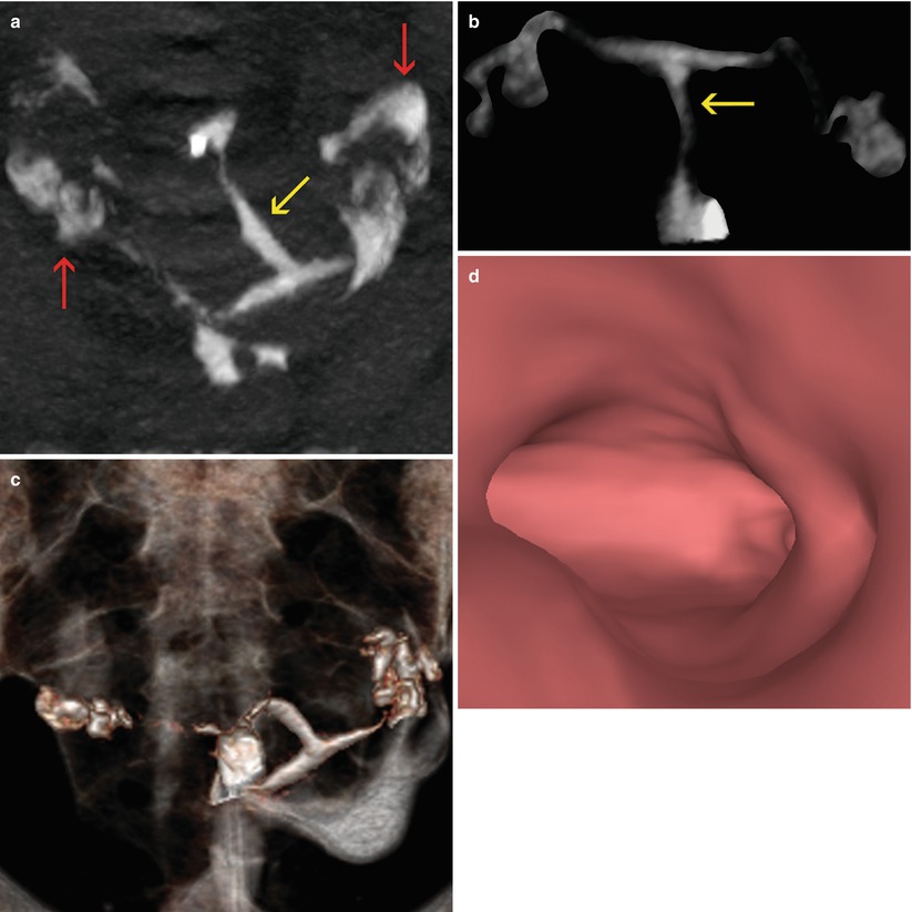
Agenesia of Müllerian Conducts

Рисунок 8.1
Radiology Key. Врожденные аномалии матки.
https://radiologykey.com/congenital-uterine-anomalies-2/

Рисунок 8.1
Unicornuate Uterus
Рисунок 8.2
Рисунок 8.3
Рисунок 8.4
Рисунок 8.5
Рисунок 8.6
Рисунок 8.7
Рисунок 8.8
Uterus didelphys with endometrial polyps
Bicornuate Uterus
Рисунок 8.9
Рисунок 8.10
Рисунок 8.11
https://radiologykey.com/uterine-wall-pathology/
Рисунок 7.1
Рисунок 7.2.
Рисунок 7.3
7.4.
Рисунок 7.5
Рисунок 7.6
Рисунок 7.7
Рисунок 7.8
Патология полости матки.
https://radiologykey.com/pathology-of-the-uterine-cavity/
Рисунок 6.1
Рисунок 6.2
Рис. 6.3.
Synechiae in uterine fundus adjacent to left horn. (a) Coronal maximum intensity projection image which shows a lineal image (arrow) at the level of the uterine fundus compatible with synechiae. (b) 3D volume rendering image which illustrates similar findings (arrow). (c) Virtual endoscopy image of the uterine fundus which exhibits a lineal band that joins the anterior and posterior walls of the uterus (synechiae) (arrow)
6.4.
Рисунок 6.5
Рисунок 6.6
Рисунок 6.7
Инфекции эндометрия.
Гипоплазия эндометрия.
Рис. 6.13.
Гиперплазия эндометрия.
Рис. 6.16.
6.17
Эндометриальные полипы
Рисунок 6.19
Рисунок 6.20
Рисунок 6.21