Radiology Key. Матка. Изменения послеоперационного периода.
https://radiologykey.com/morphological-post-treatment-changes/
Миомы.

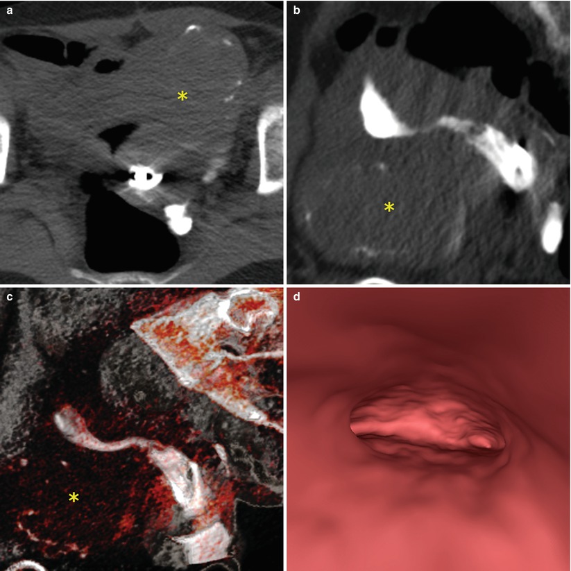
Рисунок 10.1

Рисунок 10.2


Рисунок 10.3
Radiology Key. Матка. Изменения послеоперационного периода.
https://radiologykey.com/morphological-post-treatment-changes/
Миомы.


Рисунок 10.5
Рисунок 10.7
Рисунок 10.8
Uterine Malformations
Fig. 10.9
VHSG study showing a partial septate retroverted uterus. A normal external configuration of the uterus is appreciated (yellow arrows) as well as the septum which divides the endometrial cavity (red arrows). (a) Coronal maximum intensity projection image. (b, c) Coronal 3D volume rendering images. (d) Virtual endoscopy image
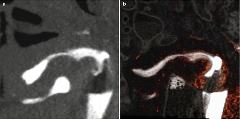
Control VHSG study after the hysteroscopy treatment. The removal of the uterine septum and the presence of a small indentation at the level of the fundus, which corresponds to the normal post-surgical septum remnant can be appreciated (arrows). (a) Coronal maximum intensity projection image. (b, c) Coronal 3D volume rendering images. (d) Virtual endoscopy image
VHSG study showing a retroverted bicornuate uterus. The concave external configuration (arrows) and the divergence of the uterine horns can be appreciated. (a) 5-mm coronal multiplanar reconstruction image. (b) Coronal maximum intensity projection image. (c) Coronal 3D volume rendering image. (d) Virtual endoscopy image
Control VHSG study after uterine repair of a bicornuate uterus. A broader endometrial cavity can be appreciated. (a) 5-mm coronal multiplanar reconstruction image. (b) Coronal maximum intensity projection image. (c) Coronal 3D volume rendering image. (d) Virtual endoscopy image
Синехии.
https://radiologykey.com/normal-radiological-anatomy/
Радиологическая анатомия матки.
Рисунок 4.1.
Положение матки
Рисунок 4.2.
Рисунок 4.3
Рисунок 4.5
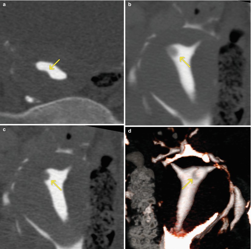
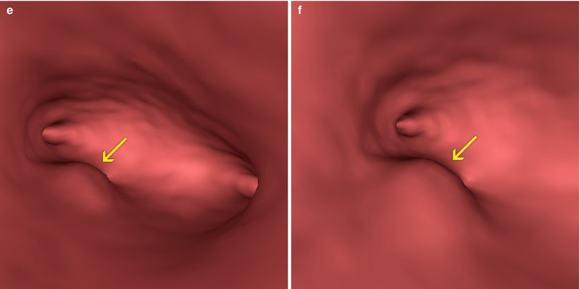
Uterine isthmus (arrow). Coronal maximum intensity projection image
Тело и дно матки.
Туберкулезная патология.
https://radiologykey.com/tubal-pathology/
Рисунок 9.1
Рисунок 9.2
Рисунок 9.3
Рисунок 9.4
Рисунок 9.5
Рисунок 9.6
Рисунок 9.7
Рисунок 9.8
Протокол исследования и интерпретация.
https://radiologykey.com/study-and-interpretation-protocol/
Патология шейки матки.
https://radiologykey.com/cervical-pathology/
Рисунок 5.1
Рис. 5.2.
Рисунок 5.3
Рисунок 5.4.
Синехии.
Рисунок 5.5
Рисунок 5.6
Рисунок 5.7
Рисунок 5.8
Thick Folds (толстые складки).
Цервикальные полипы.
Рисунок 5.11
Рисунок 5.13
Дивертикулы.
Рисунок 5.15
Кесарево сечение, "шрам".
Патология яичников.
https://radiologykey.com/incidental-findings/