A 24-year-old woman with medial foot pain following injury. (A) An anteroposterior radiograph shows a type II accessory navicular (arrow) exhibiting an articulation with the navicular bone that simulates a fracture. (B) Sagittal inversion recovery image of the mid-foot shows a type II accessory navicular that articulates with the navicular bone, with associated bone marrow edema and cystic changes around both sites of the articulation (arrow).
Figure 11.
A 39-year-old man with persistent medial foot pain and a history of prior trauma to the foot and ankle. An axial CT image shows a type II accessory navicular (arrow) articulating with the medial aspect of the navicular bone, with irregular articulating surfaces and osteophytes.


+1
С уважением. Ильич.
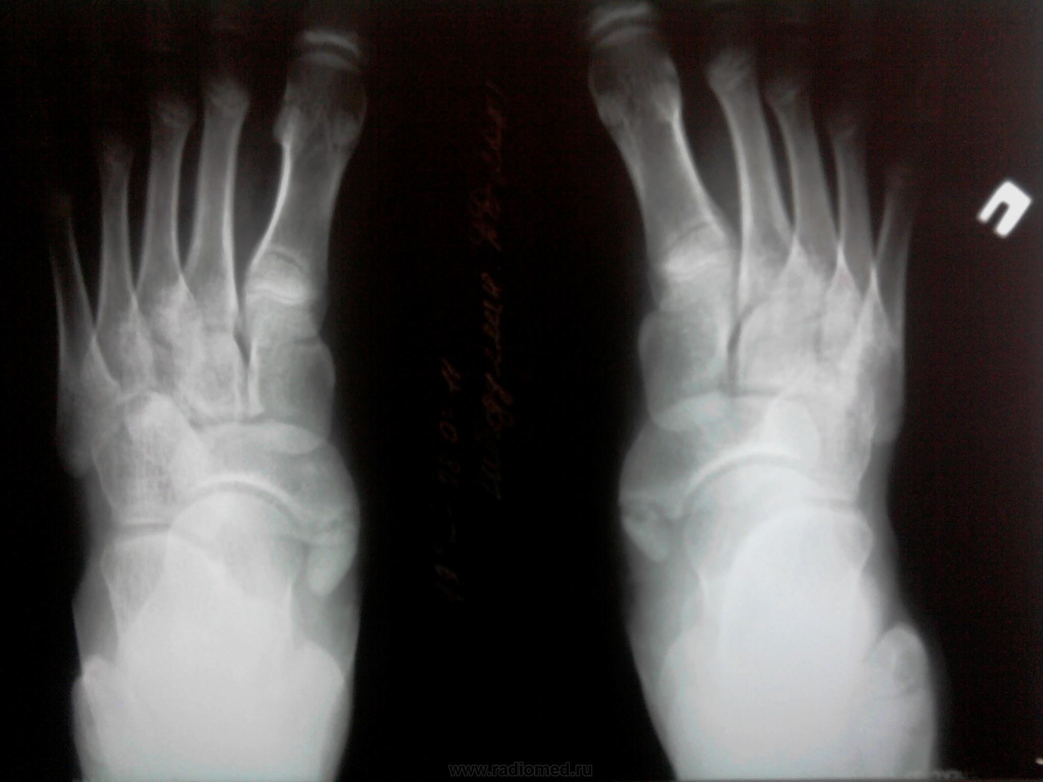
Это считается вариантом нормы?
Medicus curat, natura sanat
встреается достаточно часто.
Похожие случаи
Figure 10.
A 24-year-old woman with medial foot pain following injury. (A) An anteroposterior radiograph shows a type II accessory navicular (arrow) exhibiting an articulation with the navicular bone that simulates a fracture. (B) Sagittal inversion recovery image of the mid-foot shows a type II accessory navicular that articulates with the navicular bone, with associated bone marrow edema and cystic changes around both sites of the articulation (arrow).
Figure 11.
A 39-year-old man with persistent medial foot pain and a history of prior trauma to the foot and ankle. An axial CT image shows a type II accessory navicular (arrow) articulating with the medial aspect of the navicular bone, with irregular articulating surfaces and osteophytes.
Medicus curat, natura sanat