
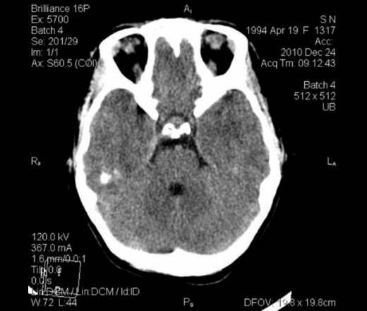
Сахарный диабет 1-го типа, поступила в сопоре, постепенно загрузилась до комы. Сахара стабилизированы, кетоацидоза нет. Тесты на наркотики положительны. Лейкоцитоз в крови. На КТ пансинусит (кроме лобных - аплазия), мастоидит, аденоиды 2-й степени.
Вопрос к коллегам: правомочно ли в этом случае писать признаки набухания вещества мозга (не отёк, а повышенное кровенаполнение)?


Реконструкции:


 Реконструкция воздушных путей


А плотность содержимого в пазухах какая? Все-таки я сторонник западного подхода к оценке состояния пазух: "Sinusitis is a clinical diagnosis. Sinus images frequently bear no relationship to the patient's symptoms. Синусит-это клинический диагноз (!!!). То что мы иногда видим в синусах, зачастую не имеет никакого отношения к симпомам которые испытывает исследуемый нами пациент" (озвучил д-р Марио).
Андрей Юрьевич
25-30 ед.Н. Я считаю, что на момент исследования кома к плотности пазух отношение не имела. Накануне введено 6 литров инфузии+метаболические дела+отравление какими-то таблетками, давшими положительный результат теста на наркотики.
https://www.youtube.com/channel/UCBGxoBUOqUT_bFhSeUgtWEw
правомочно ли в этом случае писать признаки набухания вещества мозга (не отёк, а повышенное кровенаполнение)?
https://www.youtube.com/channel/UCBGxoBUOqUT_bFhSeUgtWEw
И Вы абсолютно правы. Правда, при этом дали заключение о синусите
Андрей Юрьевич
А на основании чего? Изучения истории болезни? Описания КТ-признаков такого состояния не встречал.
Андрей Юрьевич
Тут просто показательны пазухи. А заключение включало пансинусит и мастоидит справа как сопутствующие к набуханию мозга. Уверенности, что именно термин "набухание" верен и нет. Плотность вещества не снижена, значит, не отёк... На мой взгляд, желудочки явно сужены, конвекситально местами просто не визуализируются борозды.
https://www.youtube.com/channel/UCBGxoBUOqUT_bFhSeUgtWEw
1. Пропагандирую низкопоклонство перед Западом, поэтому настороженно отношусь к попутным КТ-заключениям "Синусит" (цитату указал выше). Это же касается и мастоидита, который еще более серьезное ЛОР заболевание, требующего активного (возможно оперативного) ЛОР-лечения.
2. Повторюсь: термин "набухание" Вы сами придумали, или где-то прочитали? КТ-картина соответствует возрастной норме: "узкое - не широкое", мозгов еще слишком много.
Андрей Юрьевич
Термин "набухание головного мозга" есть такой, но по-моему, это патологоанатомический термин (как помню со студенчества), отек - это жидкость в межклеточном пространстве, а набухание - это уже в клетках, то есть набухание это хуже, чем отек.
https://www.instagram.com/pediatricradiology/
1. По Вашему, уровень жидкости в ячейках сосцевидного отростка справа имеет другое название, кроме мастоидита? Я определила именно так. В синусах жидкость гнойной плотности - писать патологическое содержимое, значит вводить клиницистов в заблуждение о природе жидкости - необходима пункция!
2. Придумывать тут нечего. "Рентгеновская компьютерная томография", Руководство для врачей под редакцией Труфанова и Рудя, глава "КТ-диагностика повреждений черепа и головного мозга", стр. 136 : " При КТ дифузное аксональное повреждение представлено общим увеличением объема мозга различной степени вследствие отёка, набухания (гиперемии) или генерализованного отёка" . Глава "КТ-диагностика инфекционных...заболеваний головного мозга", стр.181 : "Менингит. ... Возможно развитие диффузного набухания мозга"
Кроме того, при инсультах, например, одним из самых ранних признаков является сглаженность борозд и утрата ребристости островка, свидетельствующие как раз о гиперемии (см. презентацию о ранних признаках инсультов на "Файлах"). Да и не только "ранние" признаки - в любом пособии по КТ-семиотике инсультов, энцефалитов есть указание на сглаженность борозд - а чем же, по-Вашему, это обусловлено? Причём, гиперемия, т.е. повышенное кровенаполнение, характерно для многих процессов, как метаболических, так и воспалительных, а также при нарушении венозного оттока любого генеза. Все виды отёка с понижением плотности развиваются уже позже. Гиперемия и отёк являются признаками гипоксии мозга.
Ещё никто не отменял значение вентрикуло-краниальных индексов, у данной пациентки они явно ниже нормы, но при этом нет гиподенсности вещества мозга и сохранена дифференцировка между серым и белым веществом, а значит, это не отёк, а полнокровие. И часть борозд сглажена - от этого тоже трудно отмахнуться.
Но судить в данном случае мне довольно затруднительно по одной причине - нет КТ до комы. И вполне вероятно, что у девочки такие узкие желудочки от природы. Привязку к клинике я всегда учитываю (было бы глупо от неё отмахиваться). Я указала на полнокровие головного мозга у пациентки с риском отёка.
Вопрос к Вам, Андрей Юрьевич: что бы Вы написали в заключении - норму? И указали бы лишь на патологическое содержимое всех пазух?
https://www.youtube.com/channel/UCBGxoBUOqUT_bFhSeUgtWEw
Кафедра фундаментальной и клинической неврологии РГМУ, НИИ инсульта РГМУ:
"Синдром диффузного набухания мозга.
Диффузное набухание мозга проявляется генерализованным и относительно равномерным положительным объемным воздействием на ликворные прострнства: симптом щелевидных желудочков, сужение цистерн, борозд и субарахноидальных щелей. Набухание мозга связано с увеличением его кровенаполнения, что часто сочетается с отеком мозгового вещества."
https://www.youtube.com/channel/UCBGxoBUOqUT_bFhSeUgtWEw
Топик интересный. Я не силён в русских терминах, но. Выделяют два типа отёка: вазогенный и цитотоксический (внутриклеточный):
Understanding the Pathophysiology and Treatment of Brain Edema: Types of Edema and Implications for TreatmentTypes of Edema and Implications for Treatment
By definition, edema is an abnormal accumulation of fluid within the brain parenchyma; it is subdivided into vasogenic and cytotoxic types.[84] Other labels of edema such as osmotic, interstitial (hydrostatic), or hyperemic refer to etiology rather than physical location.[176]
-Vasogenic edema is defined as fluid originating from blood vessels that accumulates around cells.
-Cytotoxic edema is defined as fluid accumulating within cells as a result of injury. The most common cytotoxic edema occurs in cerebral ischemia. Heretofore, the edema specific to TBI has generally been considered to be of vasogenic origin, secondary to traumatic opening of the BBB.[15-17] However, both forms of edema can coexist. This is a critical problem, as effective treatment will clearly depend on the major type of edema contributing to the swelling process.
In the case of vasogenic edema, protein extravasation secondary to barrier compromise was implicated in the edematous process.[91] This was in line with Klatzos' theory that a breakdown of the extracellular proteins would increase the osmotic gradient and cause more water to exude from the vessels. It was shown that protein in the extracellular space retards the clearance of fluid,[52] but no evidence to date has been put forth to substantiate that extracellular protein increases fluid entry into brain. Just as protein in the extracellular space has been shown to retard clearance, lowering the ICP enhances clearance of fluid from the brain, while steroids have negligible effect on the clearance process.[68] Mediator compounds such as bradykinin, glutamate, arachidonic acid, and leukotrienes are released upon brain injury and cause brain swelling.[13,14,36,47,59,61,80,81,96,125,146,150,161,164,167,174,175]
Cytotoxic (Cellular) brain edema
November 15, 2009 at 10:07 pm · Filed under Neurological section
The author: Professor Yasser Metwally
http://yassermetwally.com
INTRODUCTION
November 15, 2009 — Cellular edema is characterized by swelling of all the cellular elements of the brain (neurons, glia, and endothelial cells), with a concomitant reduction in the volume of the extracellular fluid space of the brain. Capillary permeability is not usually affected in the various cellular edemas. Patients so affected have a normal CSF protein and isotopic brain scan. CT does not reveal enhancement with contrast, and MRI is normal.
Cellular swelling, usually of astrocytes in the grey matter, and classically is seen following cerebral ischemia caused by cardiac arrest or minor head injury. The blood brain barrier (BBB) is intact. Intracellular edema is usually not clinically significant, and is reversible in its early phases.
There are several causes of cellular edema: hypoxia, acute hypo-osmolality of the plasma, and osmotic" disequilibrium syndromes. Hypoxia after cardiac arrest results in cerebral energy depletion. The cellular swelling is osmotically determined by the appearance of increased intracellular osmoles (especially sodium, lactate, and hydrogen ions) that induce the rapid entry of water into cells. Acute hypo-osmolality of the plasma and extracellular fluid is caused by acute dilutional hyponatremia, inappropriate secretion of antidiuretic hormone, or acute sodium depletion. The brain adapts to hyponatremia by losing intracellular osmoles, chiefly potassium, thereby preserving cellular volume. Osmotic disequilibrium syndromes occur with hemodialysis or diabetic ketoacidosis, in which excessive brain intracellular solutes result in excessive cellular hydration when the plasma osmolality is rapidly reduced with therapy. The precise composition of the osmotically active intracellular solutes responsible for cellular swelling in the disequilibrium syndromes that are associated with hemodialysis and diabetic ketoacidosis is not known.
Table 1. Causes of cytotoxic brain edema
CONDITION
COMMENTS
Hypoxia
Cerebral energy depletion. The cellular swelling is osmotically determined by the appearance of increased intracellular osmoles (especially sodium, lactate, and hydrogen ions) that induce the rapid entry of water into cells.
Acute hypo-osmolality of the plasma and extracellular fluid
Caused by acute dilutional hyponatremia, inappropriate secretion of antidiuretic hormone, or acute sodium depletion, The brain adapts to hyponatremia by losing intracellular osmoles, chiefly potassium, thereby preserving cellular volume.
Osmotic disequilibrium syndromes occur with hemodialysis or diabetic ketoacidosis.
Excessive brain intracellular solutes result in excessive cellular hydration when the plasma osmolality is rapidly reduced with therapy. (In uremia, the intracellular solutes presumably include a number of organic acids, which have been recovered in the dialysis bath. In diabetic ketoacidosis, the intracellular solutes include glucose and ketone bodies; however, there are also unidentified, osmotically active, intracellular solutes, termed idiogenic osmoles that favor cellular swelling.
In uremia, the intracellular solutes presumably include a number of organic acids, which have been recovered in the dialysis bath. In diabetic ketoacidosis, the intracellular solutes include glucose and ketone bodies; however, there are also unidentified, osmotically active, intracellular solutes, termed idiogenic osmoles that favor cellular swelling. Increased intracellular osmolality in excess of the plasma level not only causes cellular swelling but also is responsible for complex changes in brain metabolism affecting the concentrations of the neurotransmitter amino acids, ammonia, and other metabolites, which in turn have profound effects on brain function.
Major changes in cerebral function occur with the cellular edemas, including stupor, coma, EEG changes and asterixis, myoclonus, and focal or generalized seizures. The encephalopathy is often severe with acute hypo- osmolality but, in more chronic state’s of hypo-osmolality of the same severity, neurologic function may be spared. Acute hypoxia causes cellular edema, which is followed by vasogenic edema as infarction develops. Vasogenic edema increases progressively for several days after an acute arterial occlusion. The delay in obtaining contrast enhancement with CT following an ischemic stroke illustrates the passage of time that is needed for defects in endothelial cell function to develop and mature.
References
Metwally, MYM: Textbook of neuroimaging, A CD-ROM publication, (Metwally, MYM editor) WEB-CD agency for electronic publication, version 10.4a October 2009 [Click to have a look at the home page]
Vasogenic edema [Full text]
3. Cerebral edema associated with nontraumatic cerebral hemorrhage [Online free full text]
Vasogenic edema
November 9, 2009 at 9:35 pm · Filed under Neurological section
The author: Professor Yasser Metwally
http://yassermetwally.com
INTRODUCTION
November 9, 2009 — Vasogenic edema is characterized by increased permeability of brain capillary endothelial cells (as consequence of vascular injury with disruption of the BBB, or due to defective endothelial lining of the newly formed blood vessels in brain neoplasms) to macromolecules, such as the plasma proteins and various other molecules, whose entry is limited by the capillary endothelial cells (blood brain barrier). Grossly, the gyri are flattened and the sulci narrowed; the white matter is moist and swollen. Microscopically, there is micro-vacuolization of the white matter, poor staining, and "halo’s" around nuclei.
Causes of vasogenic edema include trauma, tumor, abscess, hemorrhage, infarction, acute MS plaques, and cerebral contusion. It also occurs with lead encephalopathy or purulent meningitis and sinus thrombosis
Vasogenic edema is the most common type of edema associated with brain tumors, venous congestion and other causes and results from local disruption of the blood brain barrier. This leads to extravasation of protein-rich filtrate of plasma into the interstitial space, with subsequent accumulation of vascular fluid. This disruption results from loosening of the tight junctions between endothelial cells, and the neoformation of pinocytic vesicles. Once the barrier is breached, hydrostatic and osmotic forces work together to extravasate intravascular fluid. Once extravasated, fluid is retained outside the vasculature, mostly in the white matter of the brain, and within the bundles of myelinated axons of long tracts and commissural fibers. This is because axons run in parallel bundles of fibers with loose extracellular space (that offer low resistance and facilitates the extension of vasogenic edema along myelinated axons which are spreaded apart by the edema) as opposed to gray matter, which has high cell density and is enmeshed in an interwoven network of connecting fibers that offer high resistance to the formation and spread of edema. By definition, this type of edema is confined to the extracellular space. (70)
Cerebral edema may be defined broadly as a pathologic increase in the amount of total brain water content leading to an increase in brain volume 39. It occurs when plasma-like fluid enters the brain extracellular space through impaired capillary endothelial tight junctions in tumors (vasogenic edema) 40 and is a significant cause of morbidity and mortality. The molecular constituents of brain endothelial tight junctions consist of transmembrane proteins occludin, claudin 1 and 5, and junctional adhesion molecules that bind their counterparts on neighboring cells, “gluing” the cells together and creating the blood-brain barrier (BBB) 40. Intracellularly, the occludins and claudins bind to zonula occluden (ZO) 1, ZO2, and ZO3, which in turn are attached to the actin cytoskeleton 40. Normal astrocytes help to maintain a normal BBB 41, which is illustrated in Plate. 1. In high-grade tumors, the deficiency of normal astrocytes leads to defective endothelial tight junctions, resulting in BBB disruption, allowing passage of fluid into the extracellular space 40. In addition, tumor cells produce factors, such as vascular endothelial growth factor (VEGF) 42,43 and scatter factor/hepatocyte growth factor 44,45, which increase the permeability of tumor vessels by downregulation of occludin and ZO1 40,44,46,47. In addition, the membrane water channel protein, aquaporin-4 (AQP4), is upregulated around malignant brain tumors 40. AQP4-mediated transcellular water movement is important for fluid clearance in vasogenic brain edema, suggesting AQP4 activation or upregulation as a novel therapeutic target in vasogenic brain edema 40,48. High VEGF expression is reported in human anaplastic astrocytoma and glioblastoma (GBM) 49,50 meningiomas 44, and brain metastases 51. VEGF is important especially when tumors outgrow their blood supply. Hypoxia is the driving force for VEGF production in glioblastomas and the most important trigger for angiogenesis and cerebral edema formation in glioblastoma 52.
Plate 1. The BBB. Normal BBB demonstrating tight junctions between endothelial cells forming a barrier between the circulation and the brain parenchyma. Peritumoral edema formation occurs through defective endothelial junctions of an abnormal BBB. (Click to enlarge figure)
Increased capillary permeability to large molecules is the corner stone in the aetiopathogenesis of vasogenic edema. The increase in permeability is visualized when contrast enhancement is observed with CT or MRI.
The increase in permeability is visualized when contrast enhancement is observed with CT or MRI. Increased CSF protein levels are also indicative of increased endothelial permeability. MRI is more sensitive than CT in demonstrating the increased brain water and increased extracellular volume that characterize vasogenic edema. Vasogenic edema is characteristic of clinical disorders in which there is frequently a positive contrast-enhanced CT or increased signal intensity with MRI, including brain tumor, abscess, hemorrhage, infarction, and contusion. It also occurs with lead encephalopathy or purulent meningitis.
Figure 1. A, Loss of the gray-white interface with obscuration of the lentiform nucleus, loss of the insular ribbon, sulcal effacement and mass effect are seen in the left hemisphere due to vasogenic edema, B, Grossly , the gyri are flattened and the sulci narrowed; the white matter is moist and swollen. Notice uncal herniation (arrow). (Click to enlarge figure)
The functional manifestations of vasogenic edema include focal neurologic deficits, focal EEG slowing, disturbances of consciousness, and severe intracranial hypertension. In patients with brain tumor, whether primary or metastatic, the clinical signs are often caused more by the surrounding edema than by the tumor mass itself. Ultimately, these changes can lead to herniation.
Figure 2. Occipital glioblastoma surrounded by vasogenic edema involving only the white matter. (Click to enlarge figure)
Highly aggressive tumors (glioblastomas, metastatic tumours, etc.) occur at all ages; however, there is a strong trend toward increasing malignancy with age. Highly malignant tumours and rapidly growing tumours are more commonly surrounded by vasogenic tumours than more benign tumours and tumours with a lower grade of malignancy. Highly aggressive tumors are diffusely invasive tumors that typically have a destructive cellular core. Radiological signs characteristic of vasogenic brain edema is described in the following table.
Table 1. Radiological signs characteristic of vasogenic brain edema
RADIOLOGICAL SIGN
COMMENT
Contrast enhancement.
Contrast enhancement is due to break down of blood brain barrier which is the corner stone in the aetiopathogenesis of vasogenic edema. The microscopic correlate of enhancement is hypercellularity, mitotic activity, neovascularity (in brain tumours) and breakdown of blood brain barrier resulting in increased permeability of brain capillary endothelial cells to macromolecules, such as the plasma proteins and various other molecules, whose entry is limited by the capillary endothelial cells (blood brain barrier)
Diffuse low density on CT scan, diffuse MRI T1 hypointensity and diffuse MRI T2 hyperintensity with loss of the gray-white interface, obscuration of the lentiform nucleus, loss of the insular ribbon.
Obscuration of the lentiform nucleus, loss of the insular ribbon is simply due to loss of the gray-white interface.
Sulcal effacement.
Grossly , the gyri are flattened and the sulci narrowed; the white matter is moist and swollen. Microscopically, there is micro-vacuolization of the white matter, poor staining, and "halo’s" around nuclei.
Mass effect, with ventricular effacement.
Is a common cause of brain herniation.
The relationship between neuroimaging actual tumor extent is critical to the use of these studies in diagnosis and treatment design. In general three zones are identified in malignant brain tumours (1) A central zone (hypointense on the MRI T1 images, hyperintense on the MRI T2 images and hypodense on CT scan) (2) A peripheral enhanced rim with multiple enhanced mural nodules and (3) An ill-defined diffuse large zone surrounding the first two zones. (hypointense on the T1 images, hyperintense on the T2 images and hypodense on CT scan). The first zone corresponds to the necrotic tumour tissues, the microscopic correlate of enhancement is hypercellularity, mitotic activity, and neovascularity with breakdown of blood brain barrier resulting in increased permeability of brain capillary endothelial cells to macromolecules, such as the plasma proteins and various other molecules, whose entry is limited by the capillary endothelial cells (blood brain barrier), while the third zone corresponds to edema, malignant glial cell infiltrations and reactive gliosis. The surrounding zone of edema demonstrates a decreasing gradient of infiltrating tumor cells. The infiltrating tumor cells primarily follow white matter tracts, accompanied by vasogenic edema that may facilitate migration. 1,2,3,4,5 Although tumor cells may spread a great distance, typically, most are within 2 cm of the enhancing margin.6
Table 2. In general three zones are identified in malignant brain tumours
ZONE
DESCRIPTION
Central zone
Formed of necrotic tumour tissue
Intermediate contrast enhancing rim
Formed of viable tumour tissue
Peripheral diffuse zone
Formed of edema, reactive gliosis and malignant cell infiltrations
Glioblastomas characteristically send malignant cells streaming into the surrounding brain. This mode of spread is apparently facilitated by the widened extracellular spaces created through vasogenic edema.
Two types of cysts—peritumoral and intratumoral— are associated with CNS tumors. Peritumoral cysts develop within the brain or spinal cord and form at the margin of the tumor. Alternatively, intratumoral cysts develop within the tumor itself and are usually the result of intratumoral necrosis. Overall, cysts are associated with approximately 10% of benign, malignant, and metastatic tumors of the CNS. They are most frequently associated with hemangioblastomas (83%), cerebellar astrocytomas (77%), and cerebral astrocytomas (29%). The presence of peritumoral cysts can lead to significant neurological impairment due to mass effect and increased intracranial pressure. Based on advances in imaging, histological, and molecular techniques, insight into the mechanism behind peritumoral cyst formation has been provided, and evidence indicates that peritumoral edema precedes and underlies the propagation of these cysts.
Peritumoral cysts (those arising immediately adjacent to the tumor mass) are frequently associated with benign and malignant tumors of the brain and spinal cord (syringomyelia). The cystic component of central nervous system (CNS) tumors and associated peritumoral cysts are often the cause of clinical symptoms. Because of the common occurrence of peritumoral cysts with CNS neoplasms and the morbidity associated with them, advanced imaging, histological, and molecular techniques have been used to determine the mechanism underlying cyst formation and propagation. Based on evidence from such studies, edema appears to be a common precursor to peritumoral cyst formation in the CNS. Mediators of vascular permeability acting locally in the tumor and/or hydrodynamic forces within abnormal tumor vasculature appear to drive fluid extravasation. When these forces overcome the ability of surrounding tissue to resorb fluid, edema and subsequent cyst formation occur. These findings support the concept that the tumor itself is the source of the edema that precedes cyst formation and that resection of tumors or medical therapies directed at decreasing their vascular permeability will result in the resolution of edema and cysts.
Let me see...
radiographia.ru
Здесь речь не идёт об отёке. Я, по крайней мере, его не вижу. Я у девочки подозреваю полнокровие головного мозга.
А по поводу видов отёка в НИИ им. Склифосовского в 2009 году на учёбе нам давали такую классификацию отёков (разница в определении патогенеза и локализации, в приведенной Вами классификации они разделены, и внимание уделено первым двум видам, по локализации):
В зависимости от патогенетического механизма принято выделять несколько видов отека: вазогенный, цитотоксический, гидростатический, гипоосмотический, интерстициальный. По данным КТ отек характеризуется снижением плотности мозга, больше выраженное в белом веществе. При этом кортикомедуллярная граница становится менее отчетливой, что создает ощущение «монотонности» изображения.
https://www.youtube.com/channel/UCBGxoBUOqUT_bFhSeUgtWEw
В вашем случае, на КТ сканах что вы опубликовали, это исход или легкая степень cytotoxic, особенно учитывая анамнез.
"Osmotic disequilibrium syndromes occur with hemodialysis or diabetic ketoacidosis, in which excessive brain intracellular solutes result in excessive cellular hydration when the plasma osmolality is rapidly reduced with therapy. The precise composition of the osmotically active intracellular solutes responsible for cellular swelling in the disequilibrium syndromes that are associated with hemodialysis and diabetic ketoacidosis is not known."
Let me see...
radiographia.ru
"Все-таки я сторонник западного подхода к оценке состояния пазух: "Sinusitis is a clinical diagnosis. Sinus images frequently bear no relationship to the patient's symptoms. Синусит-это клинический диагноз (!!!)"
Согласитесь, что любой синуит имеет в своей основе морфологические изменения слизистой оболочки пазух. Эти изменения мы и регистрируем на КТ. Если больной ни на что не жалуется, то это вовсе не означает, что синуита нет. Ведь болезнь может иметь ремиттирующее или латентное течение.
Я сам сторонник взвешенного подхода к оценке изображений, но в данном случае согласиться не могу.
Отвечаю с опозданием - 2 дня не было электричества. Уже, наверное неактуально, но тем не менее. =Андрей Юрьевич: что бы Вы написали в заключении - норму?= Не видно совсем верхних/конвекситальных срезов, но скорее всего написал бы, что КТ-признаков патологических изменений в веществе головного не выявлено. ===И указали бы лишь на патологическое содержимое всех пазух?=== Да, и не только в этом случае. Когда обнаруживаю жидкость в плевральных полостях - пишу гидроторакс, а не плеврит.
Теперь по приведенным Вами цитатам. Труфанов и Рудь в описании диффузных аксональных повреждений не одиноки. Им вторят Лебедев и Крылов из Склифа http://neurosurgery.webzone.ru/magazine/1-2001/1_2001-9.htm
Извините, но больше верю западникам. А по англоязычной литературе КТ-признаки "Diffuse axonal injury, or DAI" (особенно без кровоизлияний) не могут быть с уверенностью представлены. На этом сайте уже обсуждалось, но наверное удалили.
А такие узкие ликворные пространства, как в представленном Вами случае, есть у любого допризывника без всякой комы.
С неизменным уважением к Вам,
Андрей Юрьевич
Я бы судил только по плотности мозга. Желудочки нормальные, борозды тоже не сужены. Можно, конечно, придраться к обводной цистерне, но это все имеет большую долю субъективизма.
Я понимаю, что термин "набухание мозга" по меньшей мере странен и не совсем корректен. Но в некоторых руководствах он встречается, и подчас имеется в виду полнокровие. Как бы то ни было, признаков отёка я не увидела. Благодарю за полезные ссылки, за высказанные мнения!
Контроль не сделала - не успела, пациентку перевели сегодня в областной центр с менингоэнцефалитом (ликвор+менингеальные), по прежнему в коме. Если станут известны новые данные, опровержение или подтверждение этого диагноза, непременно сообщу.
https://www.youtube.com/channel/UCBGxoBUOqUT_bFhSeUgtWEw
интересно а как пациентка будет жаловаться на синусит, если она в коме........
в сознании девочка, лечили как менингит. Пунктировали ли верхнечелюстные - не знаю. Будут новости - сообщу.
https://www.youtube.com/channel/UCBGxoBUOqUT_bFhSeUgtWEw
При передозе инсулина бывает гипогликемическая кома, клически тяжелая, головной мозг плохо переносит гипогликемию, часто заканчивается летальным исходом. На КТ= головного мозга первые 2-3 суток структура мозга видна, потом нарастает отек, теряется граница белого и серого вещества, появляется аксиальная дислокация с ущемлением - необратимые изменения.
Девочка выписана с выздоровлением. В области выставили менингит, но подробностей не знаю. Гипо не было - при поступлении сахара до 10, при переводе - 7.
https://www.youtube.com/channel/UCBGxoBUOqUT_bFhSeUgtWEw