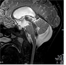
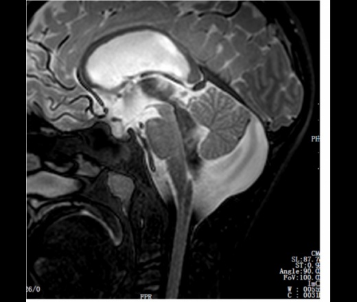
I need an expert to give an advice what is the illnes on the picture and how to treat it.
According to the neurosurgeons it is either a Blake's pouch cyst or Mega Cisterna Magna, as well as NORMAL Pressure Hydrocephalus. The doctors tell us that they cannot help him with a surgery. But the 11 years boy has many symptoms like gait disterbance, constant headache at 8,5 (0-10), nausea, stomach pain, bad apetite since late summer 2011. He was a healthy and normal boy before he got sick.
You are welcome to answer in Russian.


This is a MRI of my son, made yesterday.
Мне нужен специалист, чтобы дать совет, что это Его болезнь на картинке и как его лечить.
По словам нейрохирургов это либо чехол кисты Блейка или Mega Cisterna Magna, а также NORMAL гидроцефалия. Врачи говорят, что они не могут помочь ему с операцией. Но 11 лет мальчик имеет множество симптомов, как походка disterbance, постоянная головная боль на 8,5 (0-10), тошнота, боли в животе, плохой аппетит, так как в конце лета 2011 года. Он был здоровым и нормальным мальчиком, прежде чем он болел.
Приглашаем Вас ответить на русском языке.
Похоже на вариант мальформации Денди-Уокера. Расширена большая цистерна мозга, широко сообщается с полостью IV - го желудочка. Наверное и внутренняя гидроцефалия есть.
тоже сразу мысль про Dandy-Walker возникла
Norvejskie lekari skazali eta ne Dandy Walker.
Do you have a previous MRI to say that he was healthy? You are sure that this anomaly is not from birth?
Possible he has had it from the birth. I have no prevous MRIs when he was healthy. He has been through extensive surveys in the hospital, but nothing wrong with him. Only patological EEG and high Vitamin B12 in the blood.
.. смущает низкий сигнал от 4 и 3 желудочков.. посмотреть бы в других режимах и других проекциях..
Unfortunately I cannot publish all the MRIs, but if your are a neurosurgeon I could share the files if you send me an e-mail.
Потоковые артефакты за счёт ускорения тока ликвора.
Looks like there's an intraventricular hypertension, but we can't tell it for sure without other images from the study. At least, we need axial T2 on the level of lateral ventricles, but ideally, the whole study.
Добрый админ
I made som pictures from the case in jpg. I hope it helps. Remember he has a normal pressure hydrocefalus measured ICP with normal values.
+1
Все-таки, похоже на вариант Денди-Уокера, на мой взгляд. А говоря о гидроцефалии с нормальным давлением, Вы имеете в виду вентрикуломегалию за счет атрофического процесса или что-то другое?
врожденная мальформация - ретроцеребеллярная арахноидальная киста. Внутренняя гидроцефалия. Гипофиза маловато
...
Вариант Денди-Уокера+гидроцефалия (наверное все таки вентрикуломегалия в данном случае более корректный термин). Интересно почему "норвежские лекари" отрицают. Беспокоит также неоднородность сигнала в проекции базальных ядер.
Только подготовленный ум может рассчитывать на удачу)
Те же артефакты от ликвора.
Kakoi operacii nujna sdelat? Norvejkie lekari skazali u nemu Mega Cisterna Magna i oni ne hochit nechevo delat. Moi sin ochen bolnii... On bil sdorov i normalnii rebenok.
We are going to take him abroad in order to get treatment there. We cannot press the norwegian doctors to make surgery they do not want. Thay's why I try to get different opinions here.
Thank you everyone giving us your opinion, we are searching for right diagnose and treatment. Write in Russian, please, I understand everything but have some difficulties to write.
After ICP measuring: Mean ICP 7,8 mmHg and Mean Wave Amplitude 3,7 mm Hg.
EEG results:
19.01.12 – 30 min standard EEG with awake patient
EEG recorded under standard conditions, awake patient who is drowsy at the end of the registration. Pathological EEG with repeated bouts of rhythmic slow 3 Hz delta bilaterally but with obesity on the right side, some pointed potentials. This rhythmic activity represents suspect of epileptic activity. Not quite sure epileptic activity.
It is seen an irregular background activity 8-9 Hz alpha with symmetric distribution. It is seen repeated elements of focal rhythmic slow activity 3 Hz delta centroparietaly but also temporooccipitaly bilaterally but most prominent at the right side, mixed by slightly pointed potentials of duration 2-6 seconds and without clinical correlates. It is also seen some hints of perceived more diffuse slow activity bilaterally and may relate to the drowsiness. Some pointed potentials temporal left, but no certain of epileptic activity.
24.01.12 - 30 min standard EEG with awake patient.
It is seen irregularly 9 Hz alpha background activity with normal symmetrical distribution. Repetitive elements of focal slow activity rhythmic 3 Hz parietocentraly, partly bilaterally but most frequent on the right side, sometimes mixed with pointed/acute potentials.
It is also seen some pointed potentials but no probable or certain epileptic activity. There are seen some drowsiness and sleep-related changes.
Pathological EEG with repeated elements of focal slow activity rhythmic 3 Hz parietocentraly sometimes bilaterally but most often on the right, sometimes mixed with some pointed potentials. Beside this, irregularity 9 Hz background activity, as well as some pointed potentials, but no certain epileptic activity. The intermittent slow activity is nonspecific but seen as pathological, and somewhat more pronounced than in the previous survey.
только сегодня читал про кистозные образования мозга. Если они точно уверены что не денди-уокер, то это ретроцеребеллярная арахноидальная киста с формированием вторичной смешанной, с преобладанием внутренней, гидроцефалии (пока видимо компенсированной).
насчет тактики лечения в данном конкретном случае - то мы вам вряд ли поможем, т.к. нужна консультация нейрохирурга, специализирующегося на детях.
p.s. удачи вам!
I hope many doctors and radiologists could learn from the case of my son so not many children suffer i the future, under and missdiagnosed.
We get answer from INI Hannover that his hydrocephalus has to be treated ASAP. My son underwent ETV (Endoscopy Third Ventricle ) on 01.03.12 and now appr 2,5 months after the surgery he is much better, almost back in his healthy form.
Thinking better, almost no headache, or other pain, stronger, happier. This is a mirracle for us and tanks the doctors from INI Hannover he is a healthy boy now. He has a persistent Blake's pouch that he somehow managed to compensate for 10 years.
All the invasive examinations in the Norwegian hospital were unnessary to put the diagnose. Only MRI and symptoms are enough to consider this serios condition and I really hope my sons MRIs in this blogg could help others.
Thanks everyone who tried to advise me here in the blogg during my worst time in life!
Чем же подтвержден диагноз Blake's pouch? И, если гипертензии не было, отчего дренирующая операция помогла? Чисто по МРТ отличить Blake's pouch от мегацистерны магна проблематично. Похоже, не вся информация нам доступна.