Катенёв В.Л.
Случай из практики «Эти удивительные микрокальцинаты».
(Рентгенологическое отделение МУЗ «Красногвардейская центральная районная больница» Белгородской области)
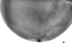
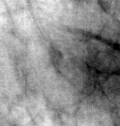
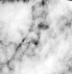
Общий фон – инволютивная железа. В верхнем наружном квадранте определяется снижение прозрачности за счёт очаговых образований местами сливного характера.
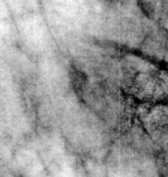
Произведено «выделение» отдельных участков железы и их изучение.
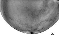
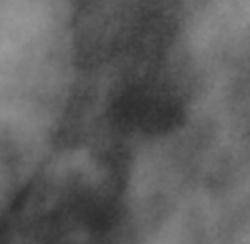
Цифровая обработка свидетельствует, что тени, которые на «первичном изображении» визуализировавшиеся, как однородные, фактически не являются таковыми. Некоторое, довольно гомогенное снижение прозрачности отдельных участков, фактически представляет собой «диффузное скопление микрокальцинатов».
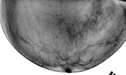
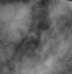
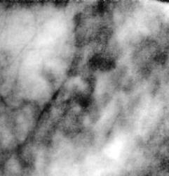
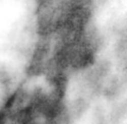
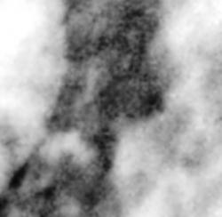
Следующие представленные изображения и последующие свидетельствуют, что субстратом довольно нечетких очаговых теней, полученных на первичном изображении, являются также микрокальцинаты.
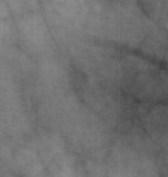
Последние три иллюстрации свидетельствуют, что отдельные микрокальцинаты, как таковые, имеют различные размеры и различную форму.
И сразу возникли следующие вопросы – недоумения:
1. Как свидетельствует классическая аналоговая рентгеномаммология, двумя основными рентгенологическими признаками рака молочной железы являются:
- непосредственное отображение опухолевого узла на рентгенограмме;
- скопление микрокальцинатов.
2. Возникает недоумение, как вообще на аналоговых маммограммах, можно было диагностировать микрокальцинаты, а главное как можно было измерить их размеры (именно в условиях практической работы в ЛПУ).


Breast calcifications:
Indicative of focally active process; often requiring biopsy. 75-80% of biopsied clusters of calcifications represent a benign process 10-30% of microcalcifications in asymptomatic patients are associated with cancers Composition: hydroxyapatite/tricalcium phosphate/calcium oxalate
Results of breast biopsies for microcalcification: (without any other mammographic findings)
Benign lesions (80%) 1. Mastopathy without proliferation 44% 2. Mastopathy with proliferation 28% 3. Fibroadenoma 4% 4. Solitary papilloma 2% 5. Miscellaneous 2%
Malignant lesions (20%) 1. Lobular carcinoma in situ in 8% no spatial relationship to LCIS 10% 2. Infiltrating carcinoma 6% 3. Ductal carcinoma in situ 4%
A positive biopsy rate of >35% is desirable goal!
Location:
intramammary Ductal microcalcifications 0.1-0.3 mm in size, irregular, sometimes mixed linear + punctate Occurrence: secretory disease, epithelial hyperplasia, atypical ductal hyperplasia, intraductal carcinoma
Lobular microcalcifications smooth round, similar in size + density Occurrence: cystic hyperplasia, adenosis, sclerosing adenosis, atypical lobular hyperplasia, lobular carcinoma in situ, cancerization of lobules (= retrograde migration of ductal carcinoma to involve lobules), ductal carcinoma obstructing egress of lobular contents N.B.: lobular and ductal microcalcifications occur frequently in fibrocystic disease + breast cancer!
Extramammary: arterial wall, duct wall, fibroadenoma, oil cyst, skin, etc.
Size: malignant calcifications usually <0.5 mm; rarely >1.0 mm Number: 5 calcifications per 1 cm2 have a low probability for malignancy
Morphology:
Benign- Smooth round calcifications: formed in dilated acini of lobules Solid/lucent-centered spheres: usually due to fat necrosis Crescent-shaped calcifications that are concave on horizontal beam lateral projection = sedimented milk of calcium at bottom of cyst Lucent-centered calcifications: around accumulated debris within ducts/in skin Solid rod-shaped calcifications/lucent-centered tubular calcifications: formed within/around normal/ectatic ducts Eggshell calcifications in rim of breast cysts Calcifications with parallel track appearance = vascular calcifications
Malignant- = calcified cellular secretions/necrotic cancer cells within ducts calcifications of vermicular form varying in size linear/branching shape
Distribution Clustered heterogeneous calcifications: adenosis, peripheral duct papilloma, hyperplasia, cancer Segmental calcifications within single duct network: suspect for multifocal cancer within lobe Regional/diffusely scattered calcifications with random distribution throughout large volumes of breast: almost always benign
Time course malignant calcifications can remain stable for >5 years!
Density Malignant Calcifications Granular calcifications = resembling fine grains of salt amorphous, dotlike/elongated, fragmented grouped very closely together irregular in form, size, and density Casting calcifications = fragmented cast of calcifications within ducts variable in size + length great variation in density within individual particles + among adjacent particles jagged irregular contour Y-shaped branching pattern clustered (>5 per focus within an area of 1 cm2)
Benign Calcifications Lobular calcifications = arise within a spherical cavity of cystic hyperplasia, sclerosing adenosis, atypical lobular hyperplasia sharply outlined, homogeneous, solid, spherical pearl-like little variation in size numerous + scattere associated with considerable fibrosis adenosis diffuse calcifications involving both breasts symmetrically periductal fibrosis diffuse/grouped calcifications + irregular borders, simulating malignant process Sedimented milk of calcium Frequency: 4% multiple, bilateral, scattered/occasionally clustered calcifications within microcysts smudge-like particles at bottom of cyst on vertical beam crescent-shaped on horizontal projection = teacup-like Plasma cell mastitis = periductal mastitis sharply marginated calcifications of uniform density = intraductal form sharply marginated hollow calcifications = periductal form
Peripheral eggshell calcifications with radiolucent lesion liponecrosis micro-/macrocystica calcificans (= fatty acids precipitate as calcium soaps at capsular surface) as calcified fat necrosis/calcified hematoma May mimic malignant calcifications! with radiopaque lesion degenerated fibroadenoma macrocyst high uniform density in periphery usually subcutaneous no associated fibrosis Papilloma solitary raspberry configuration in size of duct central/retroareolar Degenerated fibroadenoma bizarre, coarse, sharply outlined, popcorn-like very dense calcification within dense mass (= central myxoid degeneration) eggshell type calcification (= subcapsular myxoid degeneration) Arterial calcifications parallel lines of calcifications Dermal calcifications Site: sebaceous glands hollow radiolucent center polygonal shape peripheral location (may project deep within breast even on 2 views at 90° angles) linear orientation when caught in tangent same size as skin pores Proof: superficial marking technique
Metastatic calcifications Cause: hyperparathyroidism (in up to 68%)
Calcifications in Branching Tubular Opacity Ductal carcinoma in situ Atypical ductal hyperplasia Secretory disease Peripheral papillomatosis Vascular: calcified artery; Mondor disease (= thrombophlebitis of superficial vein) Fat necrosis S/P Galactography
Let me see...
radiographia.ru
Спасибо. Вечером жену посажу за перевод.
Уважаемый Dr.Mario, а это ваши личные наблюдения? Если нет, то не подскажете источник?
Ульяна Владимировна! Что Вы подразумеваете под наблюдениями?
Я имела в виду, это статья самого Доктора Марио или он взял ее из какого-либо источника и выложил на сайт для информации...